Product Detail
Product NameHistone H2A.X(Phospho Ser139) Mouse mAb
Clone No.MR4523
Host SpeciesMouse
ClonalityMonoclonal
PurificationThe antibody was affinity-purified from mouse ascites by affinity-chromatography using epitope-specific immunogen.
ApplicationsWB,IHC,IF,FC
Species ReactivityHuman,Mouse
SpecificityThis antibody detects endogenous levels of H2A.X only when phosphorylated at serine 139.
Immunogen DescSynthetic phosphopeptide corresponding to residues surrounding Ser139 of human H2A.X.
Target NameH2AFX
Other NamesH2A histone family, member X ;
H2A.X ;
H2a/x
Uniprot
P16104
Gene ID
3014
Calculated MW15KD
Sdspage MW15KD
FormulationLiquid in PBS containing 50% glycerol, 0.5% BSA and 0.02% sodium azide.
StorageStore at 4°C short term. Aliquot and store at -20°C long term. Avoid freeze/thaw cycles.
Application Details
WB 1:1000-1:2000IHC 1:200-400ICC 1:400IF 1:50-200
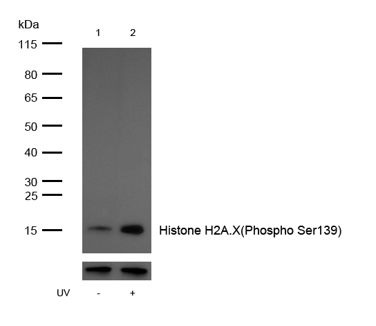
All lanes: Histone H2A.X(Phospho Ser139) Mouse mAb at 1/1k dilution Lane 1 : 293 whole cell lysates Lane 2 : 293 treated with UV for 15 minutes whole cell lysates Lysates/proteins at 20 µg per lane. Secondary All lanes : Goat Anti-Mouse IgG H&L (HRP) at 1/20000 dilution Predicted band size: 15 kDa Observed band size: 15 kDa Exposure time: 7 seconds
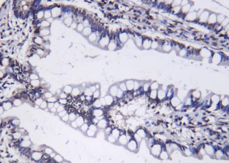
Formalin-fixed, paraffin-embedded human colon cancer tissue stained for Histone H2A.X(Phospho Ser139) using 12162 at 1/100 dilution in immunohistochemical analysis.
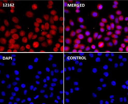
Immunocytochemistry/Immunofluorescence Histone H2A.X(Phospho Ser139) antibody (12162) ICC/IF staining of Histone H2A.X(Phospho Ser139) in Hela cells. Cells were fixed with 4% Paraformaldehyde permeabilized with 0.1% Triton X-100. Samples were incubated with 12162 at a working dilution of 1/100. The secondary antibody was Alexa Fluor® 647 goat anti mouse, used at a dilution of 1/500. The negative control is shown in bottom right hand panel - for the negative control. Nuclei were counterstained with DAPI.
Histones are basic nuclear proteins that are responsible for the nucleosome structure of the chromosomal fiber in eukaryotes. Two molecules of each of the four core histones (H2A, H2B, H3, and H4) form an octamer, around which approximately 146 bp of DNA is wrapped in repeating units, called nucleosomes. The linker histone, H1, interacts with linker DNA between nucleosomes and functions in the compaction of chromatin into higher order structures. This gene encodes a replication-independent histone that is a member of the histone H2A family, and generates two transcripts through the use of the conserved stem-loop termination motif, and the polyA addition motif. [provided by RefSeq, Oct 2015]
If you have published an article using product 12162, please notify us so that we can cite your literature.


 Yes
Yes


