Welcome to Sabbiotech!     
Email:info@signalwayantibody.com
Location: Home > Phospho Antibodies > Akt1(Phospho-Ser473) Rabbit mAb

Akt1(Phospho-Ser473) Rabbit mAb#13357

Availability: Yes
Total Amount:$
Product Detail

Product NameAkt1(Phospho-Ser473) Rabbit mAb

Clone No.SY28-05

Host SpeciesRabbit

ClonalityMonoclonal

PurificationProA affinity purified

ApplicationsWB, ICC/IF, IHC

Species ReactivityHu, Ms

Immunogen DescSynthetic phospho-peptide corresponding to residues surrounding Ser473 of human Akt1.

ConjugateUnconjugated

Other NamesAKT 1 antibody
AKT antibody
AKT1 antibody
AKT1_HUMAN antibody
MGC99656 antibody
PKB antibody
PKB-ALPHA antibody
PRKBA antibody
Protein Kinase B Alpha antibody
Protein kinase B antibody
Proto-oncogene c-Akt antibody
RAC Alpha antibody
RAC antibody
RAC-alpha serine/threonine-protein kinase antibody
RAC-PK-alpha antibody

Accession NoSwiss-Prot#:P31749

Uniprot P31749

Gene ID 207;

Calculated MWPredicted band size: 56 kDa

Sdspage MWObserved band size: 56 kDa

Formulation1*TBS (pH7.4), 1%BSA, 40%Glycerol. Preservative: 0.05% Sodium Azide.

StorageStore at -20˚C

Application Details
WB: 1:500-1:2000
ICC/IF: 1:50-1:200
IHC: 1:50-1:200
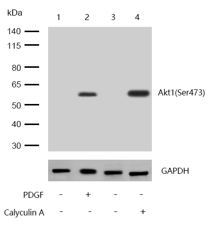
All lanes : Akt1(Phospho-Ser473) Rabbit mAb at 1/1k dilution

Lane 1 : NIH/3T3 whole cell lysates
Lane 2 : NIH/3T3 treated with 100ng/mL PDGF for 1 hour whole cell lysates
Lane 3 : MCF7 whole cell lysates
Lane 4 : MCF7 treated with 100nM Calyculin A for 30 minutes whole cell lysates

Lysates/proteins at 20 µg per lane.

Secondary
All lanes : Goat Anti-Rabbit IgG H&L (HRP) at 1/20000 dilution

Predicted band size: 56 kDa
Observed band size: 56 kDa

Exposure time: 7 seconds
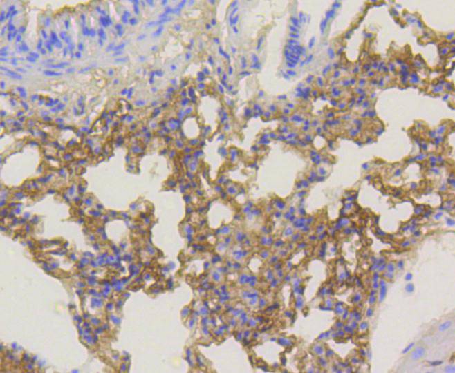
Formalin-fixed, paraffin-embedded mouse lung tissue stained for Akt1(Phospho-Ser473) using 13357 at 1/100 dilution in immunohistochemical analysis.
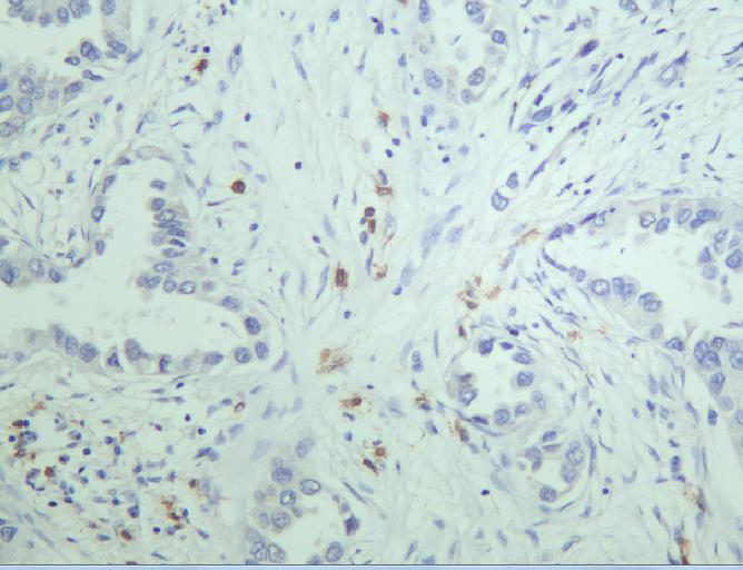
Formalin-fixed, paraffin-embedded huamn lung tissue stained for Akt1(Phospho-Ser473) using 13357 at 1/100 dilution in immunohistochemical analysis.
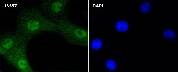
Immunocytochemistry/ Immunofluorescence Akt1(Phospho-Ser473) antibody (13357)

ICC/IF staining of Akt1(Phospho-Ser473) in NIH/3T3 cells. Cells were fixed with 4% Paraformaldehyde permeabilized with 0.1% Triton X-100.
Samples were incubated with 13357 at a working dilution of 1/100. The secondary antibody was Alexa Fluor® 488 goat anti rabbit, used at a dilution of 1/500.
Nuclei were counterstained with DAPI.
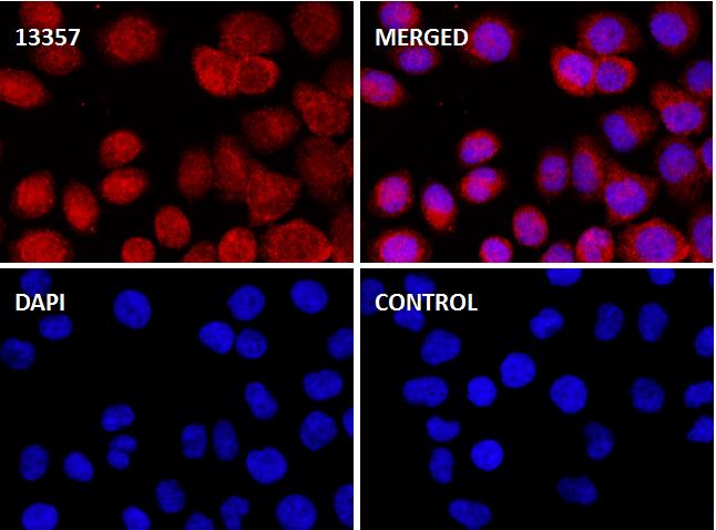
Immunocytochemistry/Immunofluorescence Akt1(Phospho-Ser473) antibody (13357)

ICC/IF staining of Akt1(Phospho-Ser473) in Hela cells. Cells were fixed with 4% Paraformaldehyde permeabilized with 0.1% Triton X-100.
Samples were incubated with 13357 at a working dilution of 1/100. The secondary antibody was Alexa Fluor® 647 goat anti rabbit, used at a dilution of 1/500.
The negative control is shown in bottom right hand panel - for the negative control.
Nuclei were counterstained with DAPI.
The serine/threonine kinase Akt family contains several members, including Akt1 (also designated PKB or RacPK), Akt2 (also designated PKBβ or RacPK-β) and Akt 3 (also designated PKBγ or thyoma viral proto-oncogene 3), which exhibit sequence homology with the protein kinase A and C families and are encoded by the c-Akt proto-oncogene. All members of the Akt family have a pleckstrin homology domain. Akt1 and Akt2 are activated by PDGF stimulation. This activation is dependent on PDGFR-β tyrosine residues 740 and 751, which bind the subunit of the phosphatidylinositol 3-kinase (PI 3-kinase) complex. Activation of Akt1 by insulin or insulin-growth factor-1(IGF-1) results in phosphorylation of both Thr 308 and Ser 473. Phosphorylation of both residues is important to generate a high level of Akt1 activity, and the phosphorylation of Thr 308 is not dependent on phosphorylation of Ser 473 in vivo. Thus, Akt proteins become phosphorylated and activated in insulin/IGF-1-stimulated cells by an upstream kinase(s). The activation of Akt1 and Akt2 is inhibited by the PI kinase inhibitor wortmannin, suggesting that the protein signals downstream of the PI kinases.

If you have published an article using product 13357, please notify us so that we can cite your literature.

et al,Downregulation of GLYAT Facilitates Tumor Growth and Metastasis and Poor Clinical Outcomes Through the PI3K/AKT/Snail Pathway in Human Breast Cancer. In Front Oncol on 2021 Apr 22 by Xin Tian, Lina Wu,et al..PMID:33968740 , (2021),
PMID: 33968740
et al,The Role of the PDGFR/PI3K/Akt Signaling Pathway in Promoting the Proliferation of Mouse Brain Astrocytes in vitro by PDGF-BB , (2019),
PMID:
et al,7?dehydrocholesterol suppresses melanoma cell proliferation and invasion via Akt1/NF?kB signaling,In Oncol Lett on 2020 Dec by Jia Liu, Feiliang Zhong, et al..PMID: 33193858 , (2020),
PMID: 33193858
et al,NVD-BM-mediated genetic biosensor triggers accumulation of 7-dehydrocholesterol and inhibits melanoma via Akt1/NF-?B signaling. In Aging (Albany NY) on 2020 Jul 25 by Jia Liu, Lei Cao, et al..PMID: 32712598 , (2020),
PMID: 32712598

NOTE

Application

  • WBWestern Blotting
  • IHCImmunohistochemistry
  • IFImmunofluorescence
  • ICCImmunocytochemistry
  • FCFlow Cytometry
  • IPImmunoprecipitation
  • EELISA
  • DBDot Blotting
  • ChIPChromatin Immunoprecipitation
  • GICAGold Immunochromatography Assay
  • NCNegative Control

Species Reactivity

  • HuHuman
  • MsMouse
  • RtRat
  • DmDrosophila melanogaster
  • CCaenorhabditis elegans
  • MkMonkey
  • RbRabbit
  • BBovine
  • DDog
  • PPig
  • HmHamster
  • ChHmChinese Hamster
  • ChkChicken
  • ShpSheep
;