Product Detail
Product NamePKC zeta(Phospho-T560) Rabbit mAb
Clone No.SN0753
Host SpeciesRabbit
ClonalityMonoclonal
PurificationProA affinity purified
ApplicationsWB,IHC
Species ReactivityHu, Ms, Rt
Immunogen DescSynthetic phospho-peptide corresponding to residues surrounding Thr560 of human PKC zeta.
ConjugateUnconjugated
Other Names14-3-3-zetaisoform antibody
AI098070 antibody
aPKCzeta antibody
C80388 antibody
EC 2.7.11.13 antibody
KPCZ_HUMAN antibody
nPKC zeta antibody
nPKC-zeta antibody
OTTHUMP00000001368 antibody
OTTHUMP00000044160 antibody
PKC 2 antibody
PKC ZETA antibody
PKC2 antibody
Pkcz antibody
PKCZETA antibody
PKM-zeta, included antibody
PRKCZ antibody
Protein kinase C zeta antibody
Protein kinase C zeta form antibody
Protein kinase C zeta type antibody
r14-3-3 antibody
R74924 antibody
zetaPKC antibody
Accession NoSwiss-Prot#:Q05513
Uniprot
Q05513
Gene ID
5590;
Calculated MWPredicted band size: 68 kDa
Sdspage MWObserved band size: 68 kDa
Formulation1*TBS (pH7.4), 1%BSA, 40%Glycerol. Preservative: 0.05% Sodium Azide.
StorageStore at -20˚C
Application Details
WB: 1:500-1:2000
IHC: 1:50-1:200
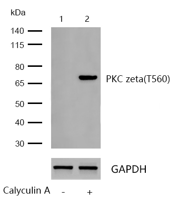
All lanes : PKC zeta(Phospho-T560) Rabbit mAb at 1/1k dilution
Lane 1 : C6 whole cell lysates
Lane 2 : C6 treated with 200nM Calyculin A for 1 hours whole cell lysate
Lysates/proteins at 20 µg per lane.
Secondary
All lanes : Goat Anti-Rabbit IgG H&L (HRP) at 1/20000 dilution
Predicted band size: 68 kDa
Observed band size: 68 kDa
Exposure time: 12 seconds
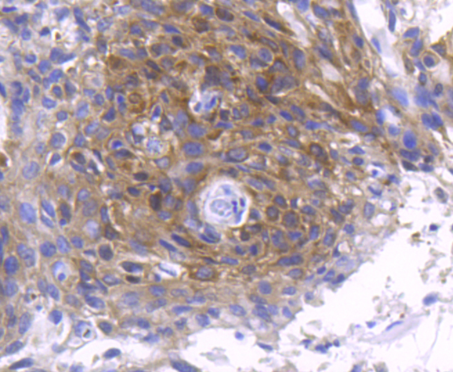
Formalin-fixed, paraffin-embedded human breast carcinoma tissue stained for PKC zeta(Phospho-T560) using 13403 at 1/100 dilution in immunohistochemical analysis.
Formalin-fixed, paraffin-embedded mouse brain tissue stained for PKC zeta(Phospho-T560) using 13403 at 1/100 dilution in immunohistochemical analysis.
Members of the protein kinase C (PKC) family play a key regulatory role in a variety of cellular functions including cell growth and differentiation, gene expression, hormone secretion and membrane function. PKCs were originally identified as serine/threonine protein kinases whose activity was dependent on calcium and phospholipids. Diacylglycerols (DAG) and tumor promoting phorbol esters bind to and activate PKC. PKCs can be subdivided into many different isoforms (α, βI, βII, γ, δ, ε, , h, θ, λ/ι, ? and n). Patterns of expression for each PKC isoform differ among tissues and PKC family members exhibit clear differences in their cofactor dependencies. For instance, the kinase activities of PKC δ and ε are independent of Ca++. On the other hand, most of the other PKC members possess phorbol ester-binding activities and kinase activities.
If you have published an article using product 13403, please notify us so that we can cite your literature.


 Yes
Yes


