
 Yes
Yes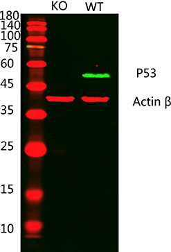
Western blot analysis of lysates from 1)p53 knockout A431 cell , 2)A431 cells, (Green) primary antibody was diluted at 1:1000, 4°over night, Dylight 800 secondary antibody was diluted at 1:10000, 37° 1hour. (Red) Actin β Monoclonal Antibody(5B7) antibody was diluted at 1:5000 as loading control, 4° over night, Dylight 680 secondary antibody was diluted at 1:10000, 37° 1hour.
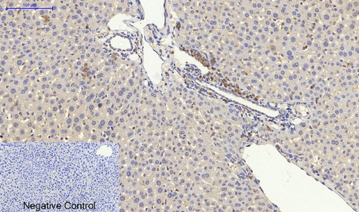
Immunohistochemical analysis of paraffin-embedded Mouse-liver tissue. 1,p53 Polyclonal Antibody was diluted at 1:200(4°C,overnight). 2, Sodium citrate pH 6.0 was used for antibody retrieval(>98°C,20min). 3,Secondary antibody was diluted at 1:200(room tempeRature, 30min). Negative control was used by secondary antibody only.
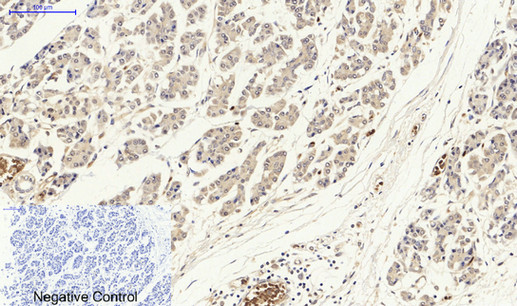
Immunohistochemical analysis of paraffin-embedded Human-lung-cancer tissue. 1,p53 Polyclonal Antibody was diluted at 1:200(4°C,overnight). 2, Sodium citrate pH 6.0 was used for antibody retrieval(>98°C,20min). 3,Secondary antibody was diluted at 1:200(room tempeRature, 30min). Negative control was used by secondary antibody only.
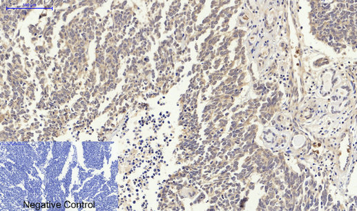
Immunohistochemical analysis of paraffin-embedded Human-stomach-cancer tissue. 1,p53 Polyclonal Antibody was diluted at 1:200(4°C,overnight). 2, Sodium citrate pH 6.0 was used for antibody retrieval(>98°C,20min). 3,Secondary antibody was diluted at 1:200(room tempeRature, 30min). Negative control was used by secondary antibody only.
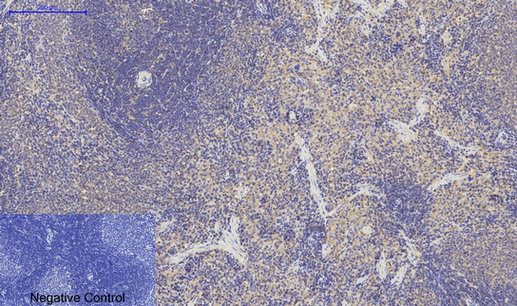
Immunohistochemical analysis of paraffin-embedded Rat-spleen tissue. 1,p53 Polyclonal Antibody was diluted at 1:200(4°C,overnight). 2, Sodium citrate pH 6.0 was used for antibody retrieval(>98°C,20min). 3,Secondary antibody was diluted at 1:200(room tempeRature, 30min). Negative control was used by secondary antibody only.
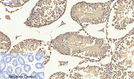
Immunohistochemical analysis of paraffin-embedded Mouse-testis tissue. 1,p53 Polyclonal Antibody was diluted at 1:200(4°C,overnight). 2, Sodium citrate pH 6.0 was used for antibody retrieval(>98°C,20min). 3,Secondary antibody was diluted at 1:200(room tempeRature, 30min). Negative control was used by secondary antibody only.
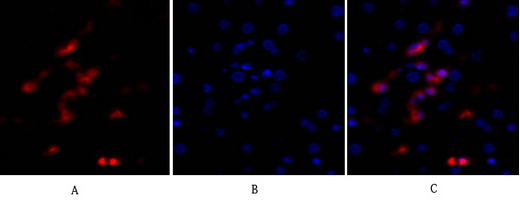
Immunofluorescence analysis of human-liver tissue. 1,p53 Polyclonal Antibody(red) was diluted at 1:200(4°C,overnight). 2, Cy3 labled Secondary antibody was diluted at 1:300(room temperature, 50min).3, Picture B: DAPI(blue) 10min. Picture A:Target. Picture B: DAPI. Picture C: merge of A+B
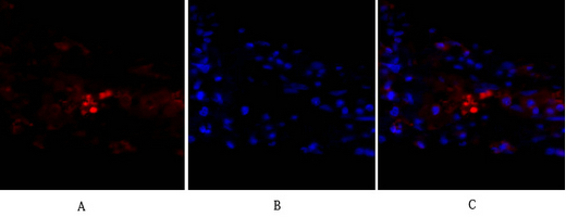
Immunofluorescence analysis of human-lung tissue. 1,p53 Polyclonal Antibody(red) was diluted at 1:200(4°C,overnight). 2, Cy3 labled Secondary antibody was diluted at 1:300(room temperature, 50min).3, Picture B: DAPI(blue) 10min. Picture A:Target. Picture B: DAPI. Picture C: merge of A+B

Immunofluorescence analysis of mouse-liver tissue. 1,p53 Polyclonal Antibody(red) was diluted at 1:200(4°C,overnight). 2, Cy3 labled Secondary antibody was diluted at 1:300(room temperature, 50min).3, Picture B: DAPI(blue) 10min. Picture A:Target. Picture B: DAPI. Picture C: merge of A+B

Figure legend: Western blot showing p53 (protein code: Tp53) expression in wild-type (WT) and Tp53 shRNA knockdown rat PC-12 cells. Beta-actin (Actb) served as the loading control.Conclusion: Since this antibody detects endogenous p53 protein in WT but not in shRNA knockdown cell lysate, this antibody is highly specific. Note: The lower band may represent the physiologically degraded p53 protein. Nonetheless, as long as these bands disappear in shRNA KD cell lysates, this antibody is still specific. This phenomenon occurs most often in polyclonal Abs, further emphasizing the importance of using KD to validate antibodies.
NOTE
Application
- WBWestern Blotting
- IHCImmunohistochemistry
- IFImmunofluorescence
- ICCImmunocytochemistry
- FCFlow Cytometry
- IPImmunoprecipitation
- EELISA
- DBDot Blotting
- ChIPChromatin Immunoprecipitation
- GICAGold Immunochromatography Assay
- NCNegative Control
Species Reactivity
- HuHuman
- MsMouse
- RtRat
- DmDrosophila melanogaster
- CCaenorhabditis elegans
- MkMonkey
- RbRabbit
- BBovine
- DDog
- PPig
- HmHamster
- ChHmChinese Hamster
- ChkChicken
- ShpSheep


