Product Detail
Product NameNFATC1 Antibody
Host SpeciesRabbit
ClonalityPolyclonal
PurificationAntibodies were purified by affinity purification using immunogen.
ApplicationsWB,IHC,IF
Species ReactivityHuman,Mouse,Rat
SpecificityThe antibody detects endogenous level of total NFATC1 protein.
Immunogen TypeRecombinant Protein
Immunogen DescRecombinant protein of human NFATC1.
Target NameNFATC1
ConjugateUnconjugated
Other NamesMGC138448; NF-ATC; NFAT2; NFATc;
Accession NoSwiss-Prot:O95644
NCBI Gene ID:4772
Uniprot
O95644
Gene ID
4772;
Sdspage MW78;101KD
Concentration1.0mg/ml
FormulationSupplied at 1.0mg/mL in phosphate buffered saline (without Mg2+ and Ca2+), pH 7.4, 150mM NaCl, 0.02% sodium azide and 50% glycerol.
StorageStore at -20˚C
Application Details
WB 1:500 - 1:3000
IHC 1:50 - 1:200
IF 1:50 - 1:200
Western blot analysis of extracts of various cell lines, using NFAT2 at 1:3000 dilution.
Immunohistochemistry of paraffin-embedded rat brain using NFAT2 at dilution of 1:100 (40x lens).
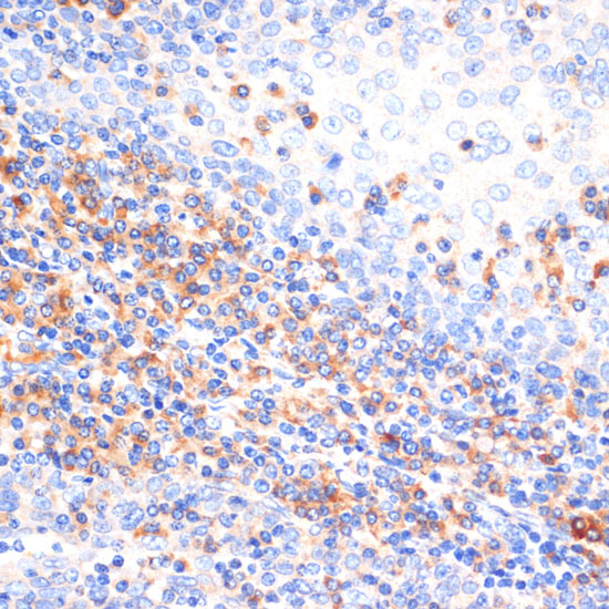
Immunohistochemistry of paraffin-embedded human tonsil using NFAT2 at dilution of 1:100 (40x lens).
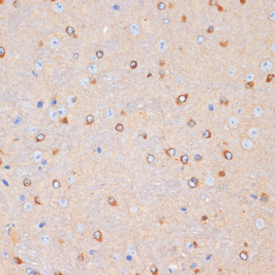
Immunohistochemistry of paraffin-embedded mouse brain using NFAT2 at dilution of 1:100 (40x lens).
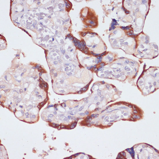
Immunohistochemistry of paraffin-embedded human placenta using NFAT2 at dilution of 1:200 (40x lens).
The NFAT (nuclear factor of activated T cells) family of proteins consists of NFAT1 (NFATc2 or NFATp), NFAT2 (NFATc1 or NFATc), NFAT3 (NFATc4), and NFAT4 (NFATc3 or NFATx). All members of this family are transcription factors with a Rel homology domain and regulate gene transcription in concert with AP-1 (Jun/Fos) to orchestrate an effective immune response (1,2). NFAT proteins are predominantly expressed in cells of the immune system, but are also expressed in skeletal muscle, keratinocytes, and adipocytes, regulating cell differentiation programs in these cells (3). In resting cells, NFAT proteins are heavily phosphorylated and localized in the cytoplasm. Increased intracellular calcium concentrations activate the calcium/calmodulin-dependent serine phosphatase calcineurin, which dephosphorylates NFAT proteins, resulting in their subsequent translocation to the nucleus (2). Termination of NFAT signaling occurs upon declining calcium concentrations and phosphorylation of NFAT by kinases such as GSK-3 or CK1 (3,4). Cyclosporin A and FK506 are immunosuppressive drugs that inhibit calcineurin and thus retain NFAT proteins in the cytoplasm (5).
If you have published an article using product 32303, please notify us so that we can cite your literature.
et al,Phytoestrogens protect joints in collagen induced arthritis by increasing IgG glycosylation and reducing osteoclast activation.In Int Immunopharmacol on 2020 Mar 12 by Du N, Song L, et al..PMID:32172207
, (2020),
PMID:
32172207
et al,Wnt7b Induced Sox11 Functions Enhance Self renewal and Osteogenic Commitment of Bone Marrow Mesenchymal Stem Cells.In Stem Cells on 2020 Apr 28. by Yu F, Wu F,et al..PMID: 32346881
, (2020),
PMID:
32346881
et al,Bioactive glass nanoparticles inhibit osteoclast differentiation and osteoporotic bone loss by activating lncRNA NRON expression in the extracellular vesicles derived from bone marrow mesenchymal stem cells. In Biomaterials on 2022 Feb 24 by Zhengyu Yang, Xiaodong Liu, et al..
, (2022),
PMID:
35220020
et al,Bioactive glass nanoparticles inhibit osteoclast differentiation and osteoporotic bone loss by activating lncRNA NRON expression in the
extracellular vesicles derived from bone marrow mesenchymal stem cells. In Biomaterials on 2022 Feb 24 by Zhengyu Yang, Xiaodong Liu, et al..PMID: 35220020
, (2022),
PMID:
35220020
et al,Increased Alleviation of Bone Destruction in Individuals with Rheumatoid Arthritis via the Coinhibition of the METTL3 and YTHDF1 Axis by the Combination of Triptolide and Medicarpin
, (2025),
PMID:

 Yes
Yes