Welcome to Sabbiotech!     
Email:info@signalwayantibody.com
Location: Home > Assay Kits > CELL-TISSUE LYSIS KIT

CELL-TISSUE LYSIS KIT#415

Availability: Inquiry (MOQ:1000 units)
Total Amount:$
Product Detail

Product NameCELL-TISSUE LYSIS KIT

ApplicationsWB

ConjugateUnconjugated

StorageStore Reagent A at -20°C Store Reagent B at room temperature

Application Details

KIT COMPONENT: Reagent A, Reagent B

DIRECTIONS:

1. Prepare the cell lysis buffer by adding 2 µL of Reagent A into 1 mL of Reagent B immediately before use. Mix well by vortexing.  2. For adherent cells, discard the medium, and wash cells twice with 

ice-cold PBS. For suspension cells, pellet down the cells by centrifugation (200g for 5 min). Resuspend the cells in 10 mL of ice-cold PBS. Pellet down the cells again, discard the PBS, and resuspend the cells in the residual buffer by pipetting.   3. For adherent cells, keep the plate/dish on ice and add 1 mL of cell lysis buffer per 5x10⁶ cells (e.g. 

add 300 µL of lysis buffer to a 35mm dish). Keep the plate/dish on ice for an additional 5 min, swirling occasionally to spread the lysis buffer. For suspension cells, add 1 mL of cell lysis buffer per 5x10⁶ cells directly to the resuspended cells. Mix by pipetting.   4. For adherent cells, after 5 min of lysis, scrap the cells off the plate/dish and collect the lysate in a centrifuge tube.  5. For both adherent and suspension cells, vortex the lysates (3 x 10 sec)and place the cells on ice for an additional 10 min to complete lysis.   6. Heat the lysates on a 95°C heat block for 5 min.  7. Cool the lysates on ice for 3 min.  8. Centrifuge the lysates at 13,000g for 5 min.   9. Measure the protein concentration using a NanoDrop spectrophotometer or an SDS_x0002_compatible protein assay method.  10. Store the cell lysates at -20°C or immediately use the lysates for further analysis. Note: For reducing gels, a final concentration of 2–5% β-mercaptoethanol or 50 mM DTT needs to be added to the lysates. The samples must be heated at 95°C for 5 min before loading.   

DIRECTIONS FOR PROTEIN EXTRACTION FROM TISSUES::

1. Grind tissues into fine particles in liquid nitrogen with a mortar and pestle.

2. Add the tissue powder into the pre_x0002_mixed IntactProteinTM lysis reagent at the ratio of 1g of tissue to 3 mL of lysis reagent. 

3. Homogenize the tissue using a homogenizer according to the manufacturer’s instructions. Tip: homogenization will heat up your sample, so always keep the tube on ice.

4. Incubate the tissue on ice for 15 min.Tip: If you deal with multiple samples, put all the samples on ice till you finish the last one. Count time from when the last sample is done. 

Incubation time longer than 15 min will not affect the quality of the extracted proteins. 

5. Centrifuge at 4°C for 10 min and transfer the supernatants into clean centrifuge tubes.

6. Follow steps 6-10 in the Directions for Protein Extraction from Cells

IntactProteinTM lysis kit preserves protein post-translational modifications (PTMs). (A-B) HeLa cells were serum starved for 16 h and stimulated with insulin (150 nM) for 5 min before harvest using IntactProteinTM lysis kit. 50 μg of total lysates were subjected to immunoblotting using anti-phospho-MTOR (Ser2448) (A) and anti-mTOR (B) antibodies, respectively. (C) HeLa cells were lysed using RIPA buffer or IntactProteinTM lysis kit. Cell lysates were blotted using anti-O-Linked β-N-acetylglucosamine (O-GlcNAc) (glycosylation) antibody.
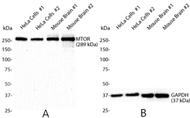
IntactProteinTM lysis kit performs equally well on protein extraction from tissues and cells. (A) HeLa cells and mouse brains (in duplicate) were lysed using IntactProteinTM lysis kit. 50 μg of total lysates were subjected to immunoblotting using anti-MTOR (289 kDa) antibody. (B) The same lysates in (A) were blotted with anti-GAPDH (37 kDa) antibody. Note that IntactProteinTM lysis kit is suitable to extract proteins of large- and small-molecular weights, and from cells and tissues.
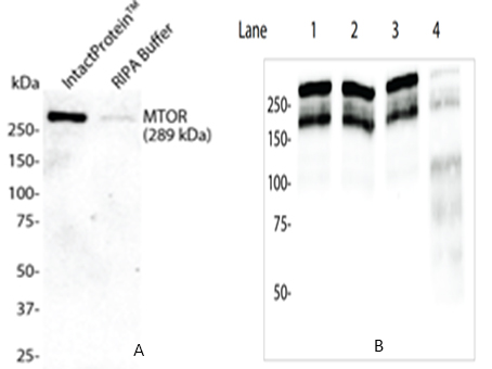
Figure 1. IntactProteinTM lysis kit shows superior performance in extracting large-sized proteins compared to RIPA buffer. (A) HeLa cells were lysed using IntactProteinTM lysis kit or RIPA buffer, and 50 μg of total lysates were subjected to immunoblotting using anti-MTOR (289 kDa)antibody. (B) HT1080 cell lysates (50 μg) were lysed on ice, processed as indicated, and blotted with anti-SMRT (160, 270 kDa) antibody. Note that sonication fragmented SMRT into smaller peptides.

One of the key factors influencing Western blot results is the extraction of proteins from cells. In practice, detergent-based buffers such as radioimmunoprecipitation assay (RIPA) buffer, along with physical disruption such as sonication, or the combination of both, have become the norm for protein extraction from cell membranes, cytoplasm, organelles, and nuclei.Although RIPA buffer (with 0.1% SDS) or its alternative such as NP-40 buffer (without SDS), has been widely used to lyse cultured mammalian cells, RIPA buffer is not as efficient at extracting large-sized proteins compared to medium- and small-sized peptides. To increase the harvest of large_x0002_sized proteins, most labs combine RIPA buffer with sonication which can physically break down DNA and thus reduce the viscosity of the lysates. Yet, sonication can break down the large-sized proteins (PLoS One, 2016; 11(1): e0148023). Additionally, to inhibit protease and phosphataseactivities, inhibitors must be added to the RIPA buffer. For example, to reduce protein degradation, protease inhibitors such as aprotinin, leupeptin, pepstatin A, and PMSF need to be added to the RIPA bufferimmediately before use. Likewise, to inhibit phosphatase activity, sodium fluoride and sodium orthovanadate must also be added.Our IntactProteinTM Cell Lysis Kit is formulated to solve these issues. It saves your time by avoiding the extra step of adding protease and phosphatase inhibitors; it can also preserve the structural and signal integrity of the cellular proteins. Further, this product is suitable for extracting all sizes of proteins from both adherent and suspension cells.


If you have published an article using product 415, please notify us so that we can cite your literature.

NOTE

Application

  • WBWestern Blotting
  • IHCImmunohistochemistry
  • IFImmunofluorescence
  • ICCImmunocytochemistry
  • FCFlow Cytometry
  • IPImmunoprecipitation
  • EELISA
  • DBDot Blotting
  • ChIPChromatin Immunoprecipitation
  • GICAGold Immunochromatography Assay
  • NCNegative Control

Species Reactivity

  • HuHuman
  • MsMouse
  • RtRat
  • DmDrosophila melanogaster
  • CCaenorhabditis elegans
  • MkMonkey
  • RbRabbit
  • BBovine
  • DDog
  • PPig
  • HmHamster
  • ChHmChinese Hamster
  • ChkChicken
  • ShpSheep
;