Product Detail
Product NameN-Cadherin Antibody
Host SpeciesRabbit
ClonalityPolyclonal
PurificationPeptide affinity purified
ApplicationsWB, ICC, IHC, FC
Species ReactivityHu, Ms
Immunogen DescSynthetic peptide (KLH-coupled) within human N-cadherin 360-420 aa.
ConjugateUnconjugated
Other NamesCADH2_HUMAN antibody
Cadherin 2 antibody
Cadherin 2 N cadherin neuronal antibody
Cadherin 2 type 1 antibody
Cadherin 2 type 1 N cadherin neuronal antibody
Cadherin 2, type 1, N-cadherin (neuronal) antibody
Cadherin-2 antibody
Cadherin2 antibody
Calcium dependent adhesion protein neuronal antibody
CD325 antibody
CD325 antigen antibody
CDH2 antibody
CDHN antibody
CDw325 antibody
CDw325 antigen antibody
N cadherin 1 antibody
N-cadherin antibody
NCAD antibody
Neural cadherin antibody
OTTHUMP00000066304 antibody
OTTHUMP00000067378 antibody
Accession NoSwiss-Prot#:P19022
Uniprot
P19022
Gene ID
1000;
Calculated MW140 kDa
Formulation1*TBS (pH7.4), 1%BSA, 40%Glycerol. Preservative: 0.05% Sodium Azide.
StorageStore at -20˚C
Application Details
WB: 1:500
IHC: 1:200
ICC: 1:200
FC: 1:50
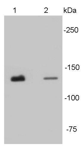
Western blot analysis of N-cadherin on different tissue lysates using anti- N-cadherin antibody at 1/500 dilution. Positive control:
Lane 1: Mouse heart
Lane2 : Human heart
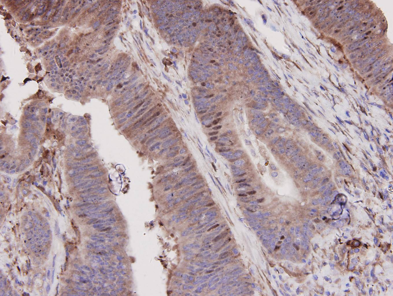
Immunohistochemical analysis of paraffin-embedded human colon carcinoma tissue using anti-N-Cadherin antibody. Counter stained with hematoxylin.
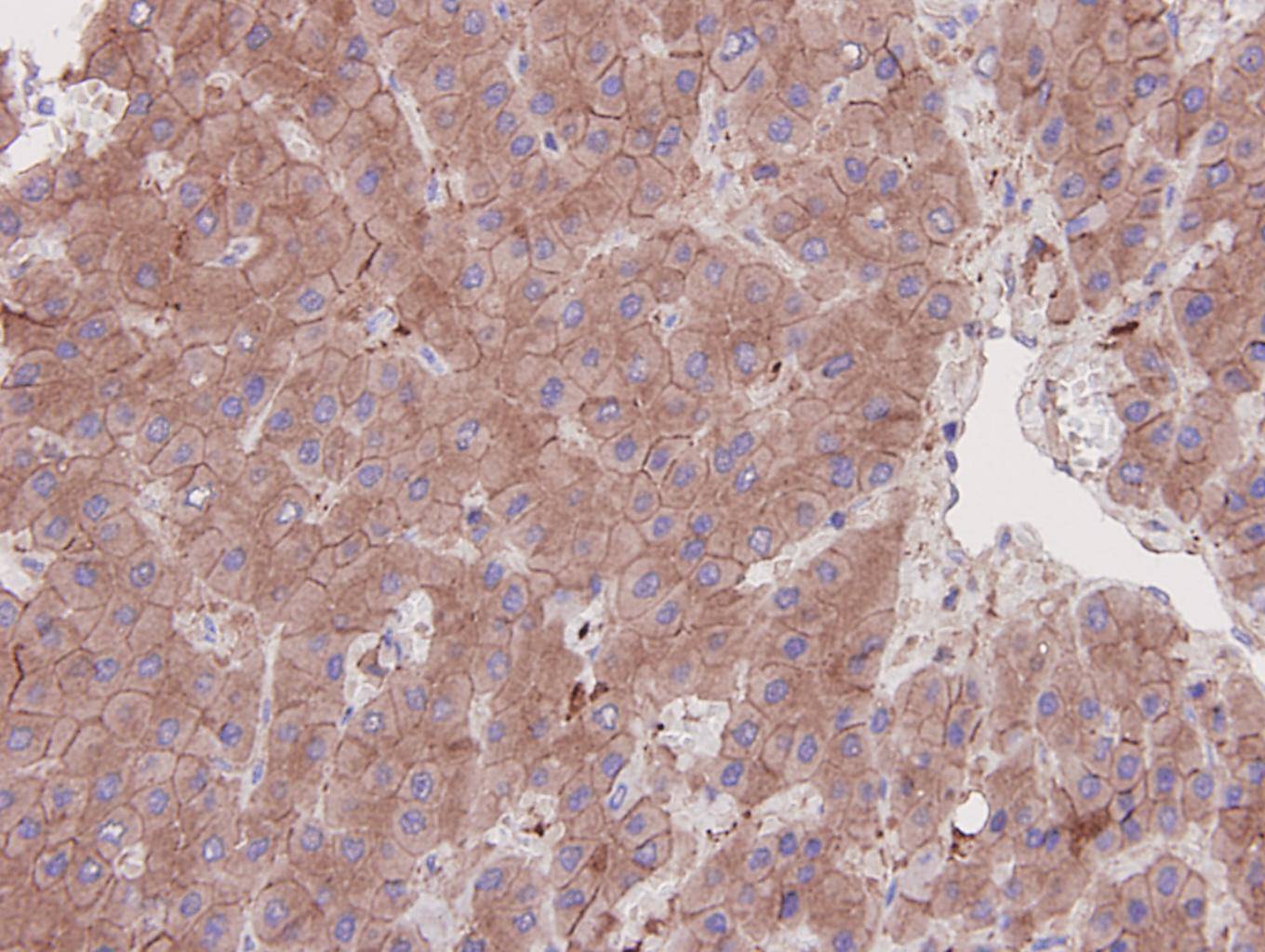
Immunohistochemical analysis of paraffin-embedded human liver carcinoma tissue using anti-N-Cadherin antibody. Counter stained with hematoxylin.
ICC staining N-Cadherin in 293 cells (red). The nuclear counter stain is DAPI (blue). Cells were Cells were fixed in paraformaldehyde, permeabilised with 0.25% Triton X100/PBS.
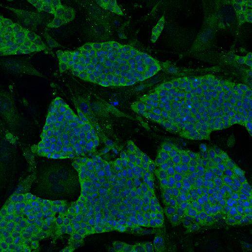
ICC staining N-Cadherin in mouse embryonic stem cells (green). The nuclear counter stain is DAPI (blue). Cells were Cells were fixed in paraformaldehyde, permeabilised with 0.25% Triton X100/PBS.
ICC staining N-Cadherin in HepG2 cells (green). The nuclear counter stain is DAPI (blue). Cells were Cells were fixed in paraformaldehyde, permeabilised with 0.25% Triton X100/PBS.
The cadherins are a large family of cell adhesion molecules involved in calcium-dependent recognition and adhesion events. The cadherin subfamily includes N-, P-, B-, and E- cadherins, and other members. Recent studies indicate that cancer cells have up-regulated N-cadherin in addition to loss of E-cadherin. N-cadherin cooperates with the FGF receptor, leading to overexpression of MMP-9 and cellular invasion. N-Cadherin is commonly found in cancer cells and provides a mechanism for transendothelial migration. When a cancer cell adheres to the endothelial cells of a blood vessel it up-regulates the src kinase pathway, which phosphorylates beta-catenins attached to both N-cadherin (this protein) and E-cadherins.
If you have published an article using product 48495, please notify us so that we can cite your literature.
et al,Neferine, a novel ROCK1-targeting inhibitor, blocks EMT process and induces apoptosis in non-small cell lung cancer. In J Cancer Res Clin Oncol on 2022 Aug 19
by Po Hu, Peng Wan, et al..PMID:35984492
, (2022),
PMID:
35984492
et al,Single-cell sequencing reveals PTX3 involvement in ovarian cancer metastasis. In J Ovarian Res on 2024 Nov 23
by Shuangyan Liu, Tianhao Wu,et al..PMID:39580424
, (2024),
PMID:
39580424


 Yes
Yes


