Welcome to Sabbiotech!     
Email:info@signalwayantibody.com
Location: Home > Recombinant Rabbit Monoclonal Antibodies > TGF-Beta 1 Antibody

TGF-Beta 1 Antibody#48569

Availability: Yes
Total Amount:$
Product Detail

Product NameTGF-Beta 1 Antibody

ClonalityMonoclonal

PurificationProtein A affinity purified.

ApplicationsWB, IHC-P, IF-Tissue

Species ReactivityHuman, Mouse, Rat

Immunogen DescSynthetic peptide (KLH-coupled) corresponding to Human TGF-Beta 1

ConjugateUnconjugated

Other NamesCartilage-inducing factor antibody
CED antibody
Differentiation inhibiting factor antibody
DPD1 antibody
LAP antibody
Latency-associated peptide antibody
Prepro transforming growth factor beta 1 antibody
TGF beta 1 antibody
TGF beta antibody
TGF beta 1 protein antibody
TGF-beta 1 protein antibody
TGF-beta-1 antibody
TGF-beta-5 antibody
TGF-beta1 antibody
TGFB antibody
Tgfb-1 antibody
tgfb1 antibody
TGFB1_HUMAN antibody
TGFbeta antibody
TGFbeta1 antibody
Transforming Growth Factor b1 antibody
Transforming Growth Factor beta 1 antibody
Transforming growth factor beta 1a antibody
transforming growth factor beta-1 antibody
transforming growth factor, beta 1 antibody
Transforming Growth Factor-?1 antibody

Accession NoSwiss-Prot#:P01137

Uniprot P01137

Gene ID 7040;

Calculated MW44 kDa

Concentration1 mg/ml

FormulationPBS (pH7.4), 0.1% BSA, 40% Glycerol. Preservative: 0.05% Sodium Azide.

StorageStore at +4℃ after thawing. Aliquot store at -20℃. Avoid repeated freeze / thaw cycles.

Application Details

WB 1:2,000
IHC-P 1:200
IF-Tissue 1:200

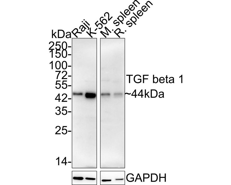
Western blot analysis of TGF beta 1 on different lysates with Rabbit anti-TGF beta 1 antibody at 1/2,000 dilution. Lane 1: Raji cell lysate (15 µg/Lane) Lane 2: K-562 cell lysate (15 µg/Lane) Lane 3: Mouse spleen tissue lysate (20 µg/Lane) Lane 4: Rat spleen tissue lysate (20 µg/Lane) Predicted band size: 44 kDa Observed band size: 44 kDa Exposure time: 3 minutes 54 seconds; 4-20% SDS-PAGE gel.
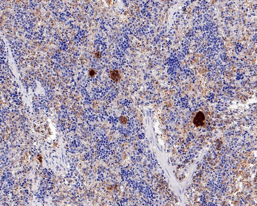
Immunohistochemical analysis of paraffin-embedded rat spleen tissue with Rabbit anti-TGF beta 1 antibody at 1/200 dilution. The section was pre-treated using heat mediated antigen retrieval with Tris-EDTA buffer (pH 9.0) for 20 minutes. The tissues were blocked in 1% BSA for 20 minutes at room temperature, washed with ddH2O and PBS, and then probed with the primary antibody at 1/200 dilution for 1 hour at room temperature. The detection was performed using an HRP conjugated compact polymer system. DAB was used as the chromogen. Tissues were counterstained with hematoxylin and mounted with DPX.
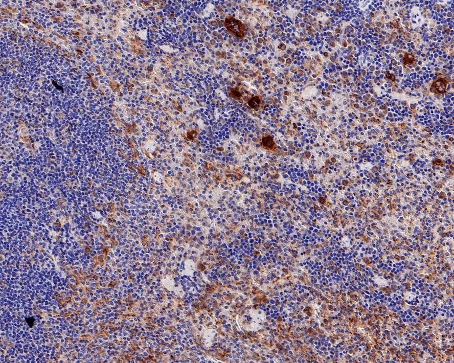
Immunohistochemical analysis of paraffin-embedded mouse spleen tissue with Rabbit anti-TGF beta 1 antibody at 1/200 dilution. The section was pre-treated using heat mediated antigen retrieval with Tris-EDTA buffer (pH 9.0) for 20 minutes. The tissues were blocked in 1% BSA for 20 minutes at room temperature, washed with ddH2O and PBS, and then probed with the primary antibody at 1/200 dilution for 1 hour at room temperature. The detection was performed using an HRP conjugated compact polymer system. DAB was used as the chromogen. Tissues were counterstained with hematoxylin and mounted with DPX.

Many cells synthesize TGFB1 and have specific receptors for it. It positively and negatively regulates many other growth factors. It plays an important role in bone remodeling as it is a potent stimulator of osteoblastic bone formation, causing chemotaxis, proliferation and differentiation in committed osteoblasts. Heterodimers of TGFB1/TGFB2 have been found in bone. Interacts with CD109 and DPT. Interacts with ASPN. Function: TGF beta 2 has suppressive effects on interleukin-2 dependent T-cell growth. Tissue specificity: TGF beta 1 is highly expressed in bone. A chromosomal aberration involving TGFB2 is found in a family with Peters anomaly.

If you have published an article using product 48569, please notify us so that we can cite your literature.

et al,Lrpap1 deficiency leads to myopia through TGF-β-induced apoptosis in zebrafish. In Cell Commun Signal on 2022 Oct 19 by Shanshan Liu, Ting Chen, et al..PMID: 36261846 , (2022),
PMID: 36261846

NOTE

Application

  • WBWestern Blotting
  • IHCImmunohistochemistry
  • IFImmunofluorescence
  • ICCImmunocytochemistry
  • FCFlow Cytometry
  • IPImmunoprecipitation
  • EELISA
  • DBDot Blotting
  • ChIPChromatin Immunoprecipitation
  • GICAGold Immunochromatography Assay
  • NCNegative Control

Species Reactivity

  • HuHuman
  • MsMouse
  • RtRat
  • DmDrosophila melanogaster
  • CCaenorhabditis elegans
  • MkMonkey
  • RbRabbit
  • BBovine
  • DDog
  • PPig
  • HmHamster
  • ChHmChinese Hamster
  • ChkChicken
  • ShpSheep
;