Product Detail
Product NameHsp70 Rabbit mAb
Clone No.SA0379
Host SpeciesRecombinant Rabbit
ClonalityMonoclonal
PurificationProA affinity purified
ApplicationsWB, ICC/IF, IHC, FC
Species ReactivityHu, Ms, Rt
Immunogen Descrecombinant protein
ConjugateUnconjugated
Other NamesDnaK type molecular chaperone HSP70 1 antibody Epididymis secretory protein Li 103 antibody FLJ54303 antibody FLJ54370 antibody FLJ54392 antibody FLJ54408 antibody FLJ75127 antibody Heat shock 70 kDa protein 1 antibody Heat shock 70 kDa protein 1/2 antibody Heat shock 70 kDa protein 1A/1B antibody Heat shock 70kDa protein 1A antibody Heat shock 70kDa protein 1B antibody Heat shock induced protein antibody HEL S 103 antibody HSP70 1 antibody HSP70 1B antibody HSP70 2 antibody HSP70-1/HSP70-2 antibody HSP70-1A antibody HSP70.1 antibody HSP70.1/HSP70.2 antibody HSP70I antibody HSP71_HUMAN antibody HSP72 antibody HSPA1 antibody HSPA1A antibody HSPA1B antibody
Accession NoSwiss-Prot#:P0DMV9
Uniprot
P0DMV9
Gene ID
3303;3304;
Calculated MW70 kDa
Formulation1*TBS (pH7.4), 1%BSA, 40%Glycerol. Preservative: 0.05% Sodium Azide.
StorageStore at -20˚C
Application Details
WB: 1:1,000-5,000
IHC: 1:50-1:200
ICC: 1:50-1:200
FC: 1:10-1:100
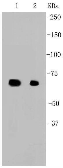
Western blot analysis of Hsp70 on different cell lysates using anti-Hsp70 antibody at 1/1,000 dilution. Positive control: Lane 1: MCF-7 Lane 2: HCT116
Immunohistochemical analysis of paraffin-embedded human breast carcinoma tissue using anti-Hsp70 antibody. Counter stained with hematoxylin.
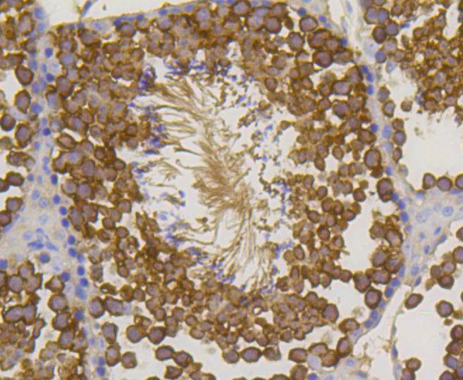
Immunohistochemical analysis of paraffin-embedded mouse testis tissue using anti-Hsp70 antibody. Counter stained with hematoxylin.
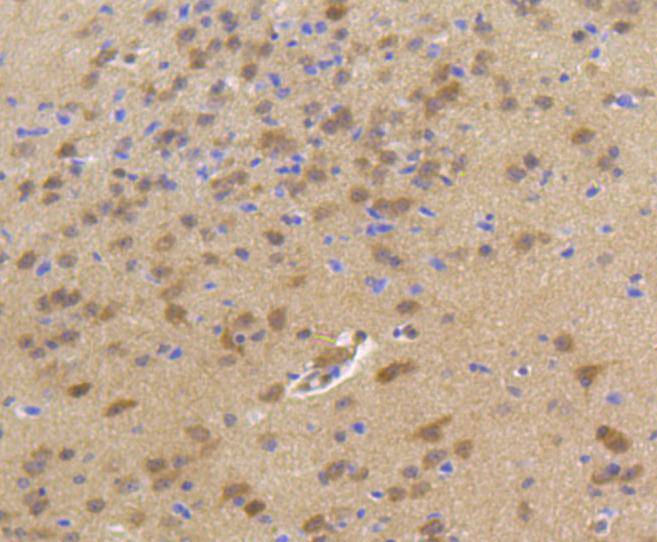
Immunohistochemical analysis of paraffin-embedded mouse brain tissue using anti-Hsp70 antibody. Counter stained with hematoxylin.
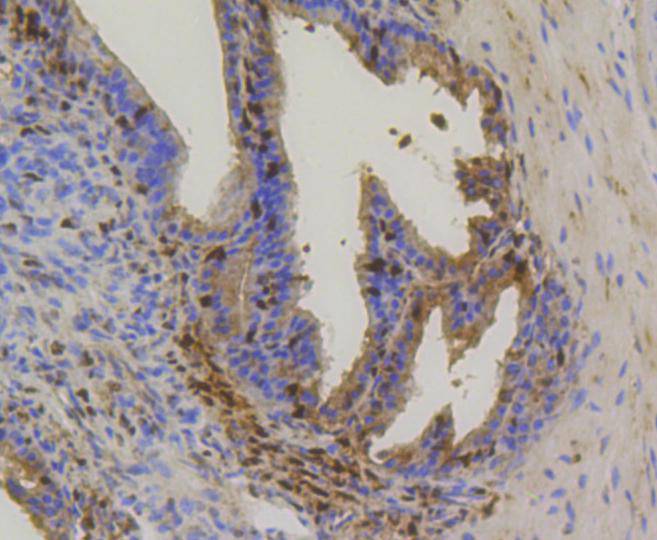
Immunohistochemical analysis of paraffin-embedded mouse prostate tissue using anti-Hsp70 antibody. Counter stained with hematoxylin.
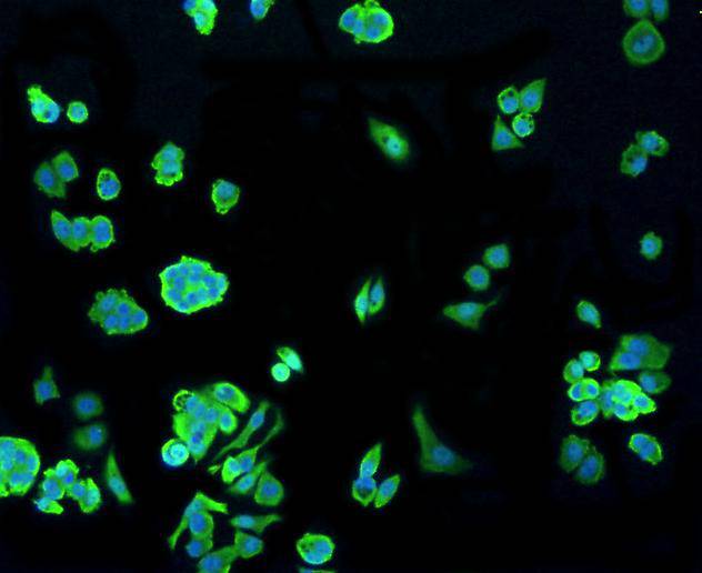
ICC staining Hsp70 in Hela cells (green). The nuclear counter stain is DAPI (blue). Cells were fixed in paraformaldehyde, permeabilised with 0.25% Triton X100/PBS.
ICC staining Hsp70 in MCF-7 cells (green). The nuclear counter stain is DAPI (blue). Cells were fixed in paraformaldehyde, permeabilised with 0.25% Triton X100/PBS.
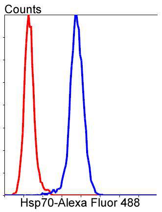
Flow cytometric analysis of Hela cells with Hsp70 antibody at 1/50 dilution (blue) compared with an unlabelled control (cells without incubation with primary antibody; red). Alexa Fluor 488-conjugated goat anti rabbit IgG was used as the secondary antibody
The 70 kilodalton heat shock proteins (Hsp70s) are a family of conserved ubiquitously expressedheat shock proteins. Proteins with similar structure exist in virtually all living organisms. The Hsp70s are an important part of the cell's machinery for protein folding, and help to protect cells from stress. When not interacting with a substrate peptide, Hsp70 is usually in an ATP bound state. Hsp70 by itself is characterized by a very weak ATPase activity, such that spontaneous hydrolysis will not occur for many minutes. As newly synthesized proteins emerge from the ribosomes, the substrate binding domain of Hsp70 recognizes sequences of hydrophobic amino acid residues, and interacts with them. This spontaneous interaction is reversible, and in the ATP bound state Hsp70 may relatively freely bind and release peptides. However, the presence of a peptide in the binding domain stimulates the ATPase activity of Hsp70, increasing its normally slow rate of ATP hydrolysis.
If you have published an article using product 48597, please notify us so that we can cite your literature.
et al,Curcumin induces apoptosis and inhibits the growth of adrenocortical carcinoma: Identification of potential candidate genes and pathways by transcriptome analysis. In Oncol Lett on 2021 Jun by Xuemei Huang, Chunfeng Liang,et al..PMID:33907586
, (2021),
PMID:
33907586




 Yes
Yes


