Product Detail
Product NameCytokeratin 19 Rabbit mAb
Clone No.SA30-06
Host SpeciesRecombinant Rabbit
ClonalityMonoclonal
PurificationProA affinity purified
ApplicationsWB, ICC/IF, IHC, FC
Species ReactivityHu, Ms
Immunogen Descrecombinant protein
ConjugateUnconjugated
Other Names40 kDa keratin intermediate filament antibody CK 19 antibody CK-19 antibody CK19 antibody Cytokeratin 19 antibody Cytokeratin-19 antibody K19 antibody K1C19_HUMAN antibody K1CS antibody Keratin 19 antibody Keratin type I 40 kD antibody Keratin type I 40kD antibody Keratin type I cytoskeletal 19 antibody Keratin, type I cytoskeletal 19 antibody Keratin, type I, 40 kd antibody Keratin-19 antibody KRT19 antibody MGC15366 antibody
Accession NoSwiss-Prot#:P08727
Uniprot
P08727
Gene ID
3880;
Calculated MW40 kDa
Formulation1*TBS (pH7.4), 1%BSA, 40%Glycerol. Preservative: 0.05% Sodium Azide.
StorageStore at -20˚C
Application Details
WB: 1:5,000-1:10,000
IHC: 1:50-1:200
ICC: 1:50-1:200
FC: 1:10-1:100
Western blot analysis of Cytokeratin 19 on MCF-7 cell lysates using anti- Cytokeratin 19 antibody at 1/10,000 dilution.
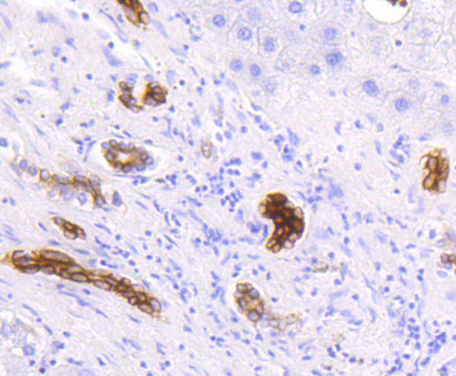
Immunohistochemical analysis of paraffin-embedded human liver tissue using anti-Cytokeratin 19 antibody. Counter stained with hematoxylin.
Immunohistochemical analysis of paraffin-embedded human breast carcinoma tissue using anti-Cytokeratin 19 antibody. Counter stained with hematoxylin.
Immunohistochemical analysis of paraffin-embedded human stomach carcinoma tissue using anti-Cytokeratin 19 antibody. Counter stained with hematoxylin.
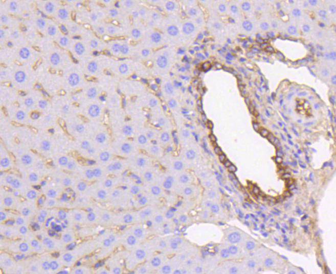
Immunohistochemical analysis of paraffin-embedded mouse liver tissue using anti-Cytokeratin 19 antibody. Counter stained with hematoxylin.
ICC staining Cytokeratin 19 in Ags cells (green). The nuclear counter stain is DAPI (blue). Cells were fixed in paraformaldehyde, permeabilised with 0.25% Triton X100/PBS.
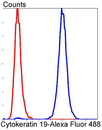
Flow cytometric analysis of MCF-7 cells with Cytokeratin 19 antibody at 1/50 dilution (blue) compared with an unlabelled control (cells without incubation with primary antibody; red). Alexa Fluor 488-conjugated goat anti-rabbit IgG was used as the secondary antibody.
Cytokeratins comprise a diverse group of intermediate filament proteins (IFPs) that are expressed as pairs in both keratinized and non-keratinized epithelial tissue. Cytokeratins play a critical role in differentiation and tissue specialization and function to maintain the overall structural integrity of epithelial cells and have been found to be useful markers of tissue differentiation, which is directly applicable to the characterization of malignant tumors. For example, many types of cancer cells express Cytokeratin 19 (CK19), an epithelial cytoskeletal protein within the suprabasal squamous epithelium. Cytokeratin 19 is a specific marker of moderate to severe dysplasia and carcinoma in situ in oral cavity squamous epithelium, and measurement of Cytokeratin 19 may be a useful marker in diagnosing hepatoma. Cytokeratin 19 fragment levels in serum have been documented as a marker for lung cancer. Clinical investigations have suggested that serum CYFRA 21-1, a fragment of Cytokeratin 19, may be among the most useful tumor markers.
If you have published an article using product 48622, please notify us so that we can cite your literature.
et al,Hepatitis B virus-related intrahepatic cholangiocarcinoma originates from hepatocytesInHepatol IntOn2023 OctbyZimin Song?#?1,?Shuirong Lin et al..PMID: 37368186
, (2023),
PMID:
37368186


 Yes
Yes


