Welcome to Sabbiotech!     
Email:info@signalwayantibody.com
Location: Home > Recombinant Rabbit Monoclonal Antibodies > Cytokeratin 17 Rabbit mAb

Cytokeratin 17 Rabbit mAb#48633

Availability: Yes
Total Amount:$
Product Detail

Product NameCytokeratin 17 Rabbit mAb

Clone No.SR45-06

Host SpeciesRecombinant Rabbit

Clonality Monoclonal

PurificationProA affinity purified

ApplicationsWB, ICC/IF, IHC, FC

Species ReactivityHu, Ms, Rt

Immunogen Descrecombinant protein

ConjugateUnconjugated

Other Names39.1 antibody
CK 17 antibody
CK-17 antibody
Cytokeratin-17 antibody
K17 antibody
K1C17_HUMAN antibody
Keratin 17 antibody
keratin 17 epitope S1 antibody
keratin 17 epitope S2 antibody
keratin 17 epitope S4 antibody
Keratin 17, type I antibody
Keratin antibody
Keratin type I cytoskeletal 17 antibody
keratin, type i cytoskeletal 17 [version 1] antibody
Keratin-17 antibody
KRT17 antibody
PC antibody
PC2 antibody
PCHC1 antibody
type I cytoskeletal 17 antibody

Accession NoSwiss-Prot#:Q04695

Uniprot Q04695

Gene ID 3872;

Calculated MW48 kDa

Formulation1*TBS (pH7.4), 1%BSA, 40%Glycerol. Preservative: 0.05% Sodium Azide.

StorageStore at -20˚C

Application Details
WB: 1:500-1:1,000
IHC: 1:50-1:200
ICC: 1:50-1:200

FC: 1:50-1:100
Immunohistochemical analysis of paraffin-embedded human tonsil tissue using anti-Cytokeratin 17 antibody. Counter stained with hematoxylin.
Immunohistochemical analysis of paraffin-embedded human breast carcinoma tissue using anti-Cytokeratin 17 antibody. Counter stained with hematoxylin.
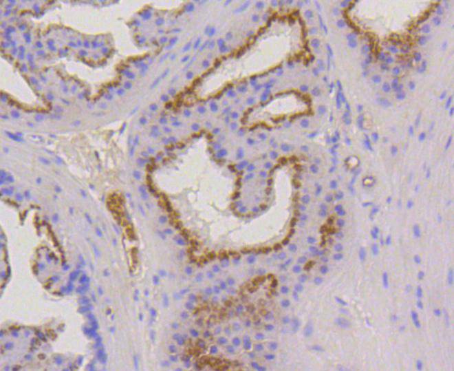
Immunohistochemical analysis of paraffin-embedded mouse prostate tissue using anti-Cytokeratin 17 antibody. Counter stained with hematoxylin.
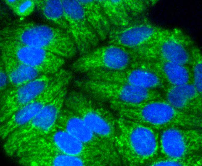
ICC staining Cytokeratin 17 in Hela cells (green). The nuclear counter stain is DAPI (blue). Cells were fixed in paraformaldehyde, permeabilised with 0.25% Triton X100/PBS.
ICC staining Cytokeratin 17 in PC-3M cells (green). The nuclear counter stain is DAPI (blue). Cells were fixed in paraformaldehyde, permeabilised with 0.25% Triton X100/PBS.
Flow cytometric analysis of Hela cells with Cytokeratin 17 antibody at 1/50 dilution (blue) compared with an unlabelled control (cells without incubation with primary antibody; red). Alexa Fluor 488-conjugated goat anti rabbit IgG was used as the secondary antibody.
Cytokeratin 17 is a member of the Cytokeratin subfamily of intermediate filament proteins (IFPs). It is unique in that it is normally expressed in the basal cells of complex epithelia but not in stratified or simple epithelia. Cytokeratin 17 contains 432 amino acids and is expressed in the nail bed, hair follicle, sebaceous glands and other epidermal appendages. Cytokeratin 17 functions to regulate cell growth and size through its interactions with the adaptor protein 14-3-3-sigma to mediate protein synthesis. Mutations in the gene encoding for Cytokeratin 17 lead to depressed protein translation and smaller sized skin keratinocytes, corresponding to decreased Akt/mTOR signaling activity. Cytokeratin 17 may be a useful marker for cervical stem cell identification, squamous cell carcinoma of the larynx, respiratory syncytial virus and transitional cell carcinomas of the human urinary tract.

If you have published an article using product 48633, please notify us so that we can cite your literature.

NOTE

Application

  • WBWestern Blotting
  • IHCImmunohistochemistry
  • IFImmunofluorescence
  • ICCImmunocytochemistry
  • FCFlow Cytometry
  • IPImmunoprecipitation
  • EELISA
  • DBDot Blotting
  • ChIPChromatin Immunoprecipitation
  • GICAGold Immunochromatography Assay
  • NCNegative Control

Species Reactivity

  • HuHuman
  • MsMouse
  • RtRat
  • DmDrosophila melanogaster
  • CCaenorhabditis elegans
  • MkMonkey
  • RbRabbit
  • BBovine
  • DDog
  • PPig
  • HmHamster
  • ChHmChinese Hamster
  • ChkChicken
  • ShpSheep
;