Product Detail
Product NameDUSP6 Rabbit mAb
Clone No.SR39-09
Host SpeciesRecombinant Rabbit
Clonality Monoclonal
PurificationProA affinity purified
ApplicationsWB, ICC/IF, IHC, IP, FC
Species ReactivityHu, Ms, Rt
Immunogen Descrecombinant protein
ConjugateUnconjugated
Other NamesDual specificity phosphatase 6 antibody
Dual specificity phosphatase 6 isoform a antibody
Dual specificity protein phosphatase 6 antibody
Dual specificity protein phosphatase PYST1 antibody
DUS6_HUMAN antibody
DUSP 6 antibody
DUSP 6a antibody
Dusp6 antibody
DUSP6a antibody
HH19 antibody
MAP kinase phosphatase 3 antibody
Mitogen activated protein kinase phosphatase 3 antibody
Mitogen-activated protein kinase phosphatase 3 antibody
MKP 3 antibody
MKP-3 antibody
MKP3 antibody
PYST 1 antibody
PYST1 antibody
Serine/threonine specific protein phosphatase antibody
Accession NoSwiss-Prot#:Q16828
Uniprot
Q16828
Gene ID
1848;
Calculated MW42 kDa
Formulation1*TBS (pH7.4), 1%BSA, 40%Glycerol. Preservative: 0.05% Sodium Azide.
StorageStore at -20˚C
Application Details
WB: 1:1,000-5,000
IHC: 1:50-1:200
ICC: 1:50-1:200
FC: 1:50-1:100
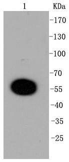
Western blot analysis of DUSP6 on mouse pancreas lysates using anti-DUSP6 antibody at 1/1,000 dilution.
Immunohistochemical analysis of paraffin-embedded human gastric carcinoma tissue using anti-DUSP6 antibody. Counter stained with hematoxylin.
Immunohistochemical analysis of paraffin-embedded human pancreas tissue using anti-DUSP6 antibody. Counter stained with hematoxylin.
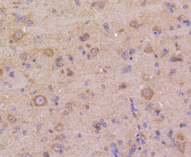
Immunohistochemical analysis of paraffin-embedded mouse brain tissue using anti-DUSP6 antibody. Counter stained with hematoxylin.
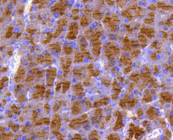
Immunohistochemical analysis of paraffin-embedded mouse pancreas tissue using anti-DUSP6 antibody. Counter stained with hematoxylin.
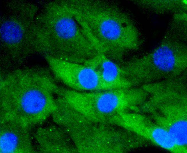
ICC staining DUSP6 in Hela cells (green). The nuclear counter stain is DAPI (blue). Cells were fixed in paraformaldehyde, permeabilised with 0.25% Triton X100/PBS.
ICC staining DUSP6 in PC-12 cells (green). The nuclear counter stain is DAPI (blue). Cells were fixed in paraformaldehyde, permeabilised with 0.25% Triton X100/PBS.
ICC staining DUSP6 in NIH/3T3 cells (green). The nuclear counter stain is DAPI (blue). Cells were fixed in paraformaldehyde, permeabilised with 0.25% Triton X100/PBS.
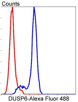
Flow cytometric analysis of NIH/3T3 cells with DUSP6 antibody at 1/50 dilution (blue) compared with an unlabelled control (cells without incubation with primary antibody; red). Alexa Fluor 488-conjugated goat anti rabbit IgG was used as the secondary antibody
Mitogen-activated protein (MAP) kinases are a large class of proteins involved in signal transduction pathways that are activated by a range of stimuli and mediate a number of physiological and pathological changes in the cell. Dual specificity phosphatases (DSPs) are a subclass of the protein tyrosine phosphatase (PTP) gene superfamily, which are selective for dephosphorylating critical phosphothreonine and phosphotyrosine residues within MAP kinases. DSP gene expression is induced by a host of growth factors and/or cellular stresses, thereby negatively regulating MAP kinase superfamily members including MAPK/ERK, SAPK/JNK and p38. The members of the dual-specificity phosphatase protein family include MKP-1/CL100 (3CH134), VHR, PAC1, MKP-2, hVH-3 (B23), hVH-5, MKP-3, MKP-X, and MKP-4. Human MKP-3 maps to chromosome 12q22-q23 and encodes a 381 amino acid protein that specifically inactivates members of the ERK family and is expressed in a variety of tissues with the highest levels in heart and pancreas.
If you have published an article using product 48635, please notify us so that we can cite your literature.






 Yes
Yes


