Product Detail
Product NamePUMA Rabbit mAb
Clone No.SR42-09
Host SpeciesRecombinant Rabbit
Clonality Monoclonal
PurificationProA affinity purified
ApplicationsWB, ICC/IF, IHC, FC
Species ReactivityHu, Ms, Rt
Immunogen Descrecombinant protein
ConjugateUnconjugated
Other NamesBBC 3 antibody
Bbc3 antibody
BBC3_HUMAN antibody
BCL 2 binding component 3 antibody
Bcl-2-binding component 3 antibody
BCL2 binding component 3 antibody
JFY 1 antibody
JFY-1 antibody
JFY1 antibody
p53 up regulated modulator of apoptosis antibody
p53 up-regulated modulator of apoptosis antibody
p53 Upregulated Modulator of Apoptosis antibody
PUMA alpha antibody
PUMA/JFY1 antibody
Accession NoSwiss-Prot#:Q9BXH1
Uniprot
Q96PG8
Gene ID
27113;
Calculated MW18 kDa
Formulation1*TBS (pH7.4), 1%BSA, 40%Glycerol. Preservative: 0.05% Sodium Azide.
StorageStore at -20˚C
Application Details
WB: 1:1,000-1:2,000
IHC: 1:50-1:200
ICC: 1:50-1:200
FC: 1:50-1:100
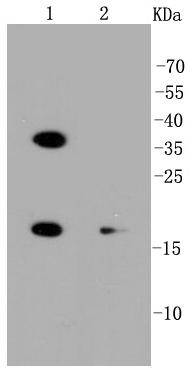
Western blot analysis of PUMA on different lysates using anti-PUMA antibody at 1/1,000 dilution. Positive control:
Lane 1: Hela
Lane 2: K562
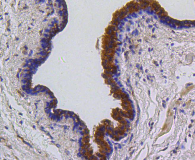
Immunohistochemical analysis of paraffin-embedded human breast carcinoma tissue using anti-PUMA antibody. Counter stained with hematoxylin.
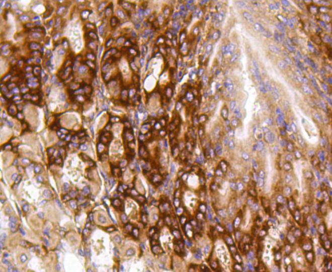
Immunohistochemical analysis of paraffin-embedded human gastric carcimnoma tissue using anti-PUMA antibody. Counter stained with hematoxylin.
Immunohistochemical analysis of paraffin-embedded mouse stomach tissue using anti-PUMA antibody. Counter stained with hematoxylin.
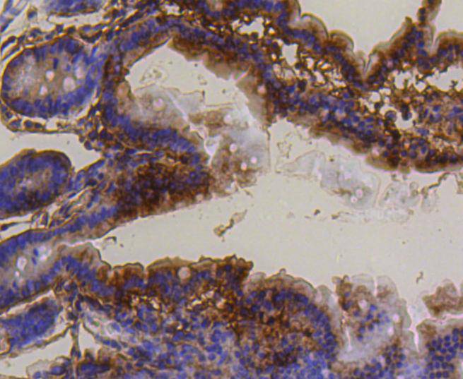
Immunohistochemical analysis of paraffin-embedded mouse small intestine tissue using anti-PUMA antibody. Counter stained with hematoxylin.
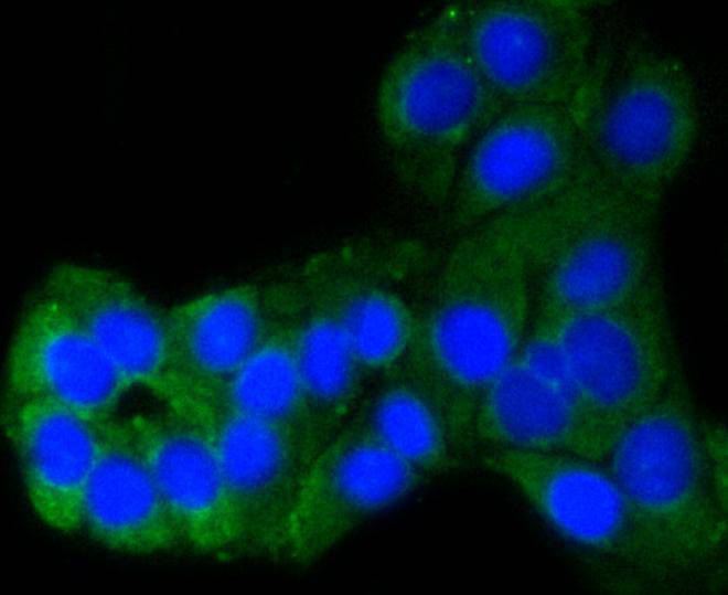
ICC staining PUMA in Hela cells (green). The nuclear counter stain is DAPI (blue). Cells were fixed in paraformaldehyde, permeabilised with 0.25% Triton X100/PBS.
ICC staining PUMA in SKOV-3 cells (green). The nuclear counter stain is DAPI (blue). Cells were fixed in paraformaldehyde, permeabilised with 0.25% Triton X100/PBS.
Flow cytometric analysis of Hela cells with PUMA antibody at 1/50 dilution (blue) compared with an unlabelled control (cells without incubation with primary antibody; red). Alexa Fluor 488-conjugated goat anti rabbit IgG was used as the secondary antibody
The expression of PUMA is regulated by the tumor suppressor p53. PUMA is involved in p53-dependent and -independent apoptosis induced by a variety of signals, and is regulated by transcription factors, not by post-translational modifications. After activation, PUMA interacts with antiapoptotic Bcl-2 family members, thus freeing Bax and/or Bak which are then able to signal apoptosis to the mitochondria. Following mitochondrial dysfunction, the caspase cascade is activated ultimately leading to cell death. Several studies have shown that PUMA function is affected or absent in cancer cells. Additionally, many human tumors contain p53 mutations, which results in no induction of PUMA, even after DNA damage induced through irradiation or chemotherapy drugs.Other cancers, which exhibit overexpression of antiapotptic Bcl-2 family proteins, counteract and overpower PUMA-induced apoptosis.
If you have published an article using product 48642, please notify us so that we can cite your literature.





 Yes
Yes


