Product Detail
Product NameCaveolin-1 Rabbit mAb
Clone No.SZ02-01
Host SpeciesRecombinant Rabbit
Clonality Monoclonal
PurificationProA affinity purified
ApplicationsWB, ICC/IF, IHC
Species ReactivityHu
Immunogen Descrecombinant protein
ConjugateUnconjugated
Other NamesBSCL3 antibody CAV antibody CAV1 antibody CAV1_HUMAN antibody caveolae protein, 22 kD antibody caveolin 1 alpha isoform antibody caveolin 1 beta isoform antibody Caveolin 1 caveolae protein 22kDa antibody Caveolin-1 antibody Caveolin1 antibody cell growth-inhibiting protein 32 antibody CGL3 antibody LCCNS antibody MSTP085 antibody OTTHUMP00000025031 antibody PPH3 antibody VIP 21 antibody VIP21 antibody
Accession NoSwiss-Prot#:Q03135
Uniprot
Q03135
Gene ID
857;
Calculated MW20 kDa
Formulation1*TBS (pH7.4), 1%BSA, 40%Glycerol. Preservative: 0.05% Sodium Azide.
StorageStore at -20˚C
Application Details
WB: 1:1,000-5,000
IHC: 1:50-1:200
ICC: 1:50-1:200
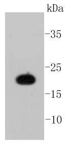
Western blot analysis of Caveolin-1 on A431 cell lysates using anti-Caveolin-1 antibody at 1/1,000 dilution.
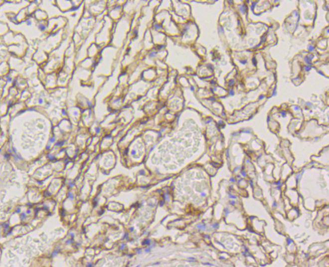
Immunohistochemical analysis of paraffin-embedded human lung tissue using anti-Caveolin-1 antibody. Counter stained with hematoxylin.
Immunohistochemical analysis of paraffin-embedded human liver tissue using anti-Caveolin-1 antibody. Counter stained with hematoxylin.
Immunohistochemical analysis of paraffin-embedded mouse lung tissue using anti-Caveolin-1 antibody. Counter stained with hematoxylin.
Immunohistochemical analysis of paraffin-embedded mouse liver tissue using anti-Caveolin-1 antibody. Counter stained with hematoxylin.
Immunohistochemical analysis of paraffin-embedded mouse heart tissue using anti-Caveolin-1 antibody. Counter stained with hematoxylin.
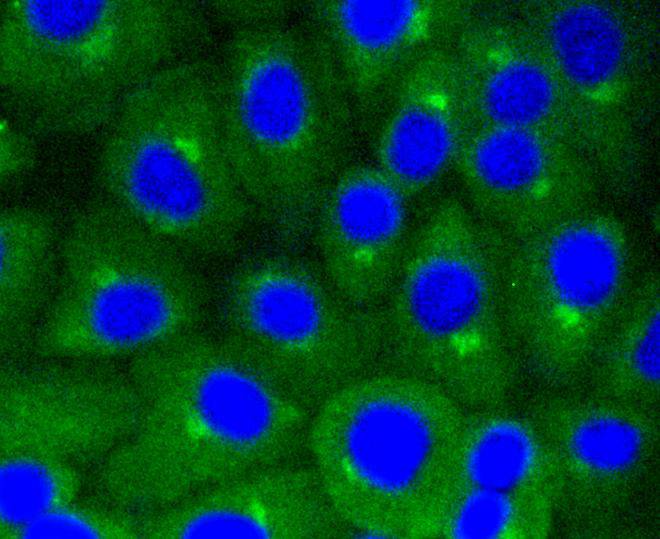
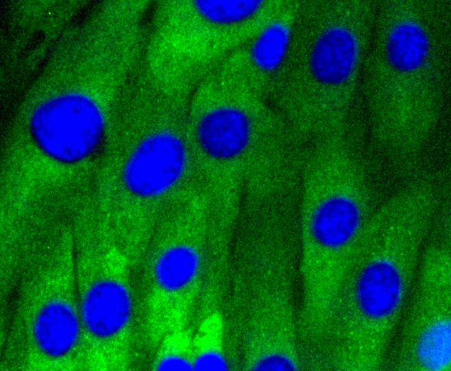
ICC staining Caveolin-1 in A549 cells (green). The nuclear counter stain is DAPI (blue). Cells were fixed in paraformaldehyde, permeabilised with 0.25% Triton X100/PBS.
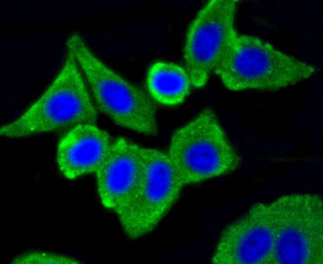
ICC staining Caveolin-1 in MCF-7 cells (green). The nuclear counter stain is DAPI (blue). Cells were fixed in paraformaldehyde, permeabilised with 0.25% Triton X100/PBS.
ICC staining Caveolin-1 in NIH/3T3 cells (green). Cells were fixed in paraformaldehyde, permeabilised with 0.25% Triton X100/PBS.
Caveolae (also known as plasmalemmal vesicles) are 50-100 nM flask-shaped membranes that represent a subcompartment of the plasma membrane. On the basis of morphological studies, caveolae have been implicated to function in the transcytosis of various macromolecules (including LDL) across capillary endothelial cells, uptake of small molecules via potocytosis and the compartmentalization of certain signaling molecules including G protein-coupled receptors. Three proteins, caveolin-1, caveolin-2 and caveolin-3, have been identified as principal components of caveolae. Two forms of caveolin-1, designated alpha and beta, share a distinct but overlapping cellular distribution and differ by an amino terminal 31 amino acid sequence which is absent from the beta isoform. Caveolin-1 shares 31% identity with caveolin-2 and 65% identity with caveolin-3 at the amino acid level. Functionally, the three proteins differ in their interactions with heterotrimeric G protein isoforms.
If you have published an article using product 48674, please notify us so that we can cite your literature.




 Yes
Yes


