
 Yes
Yes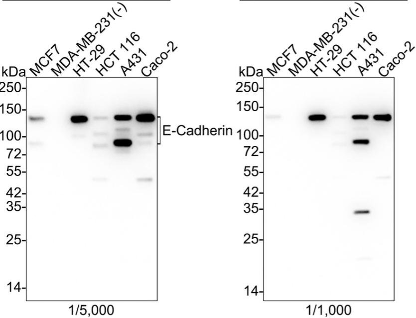
Western blot analysis of E Cadherin on different lysates with E Cadherin antibody at 1/5,000 dilution and competitor's antibody at 1/1,000 dilution.;
Lane 1: MCF7 cell lysate;
Lane 2: MDA-MB-231 cell lysate (negative);
Lane 3: HT-29 cell lysate;
Lane 4: HCT 116 cell lysate;
Lane 5: A431 cell lysate;
Lane 6: Caco-2 cell lysate;
Lysates/proteins at 20 ug/Lane.;
Predicted band size: 97 kDa;
Observed band size: 80-120 kDa
Lane 1: MCF7 cell lysate;
Lane 2: MDA-MB-231 cell lysate (negative);
Lane 3: HT-29 cell lysate;
Lane 4: HCT 116 cell lysate;
Lane 5: A431 cell lysate;
Lane 6: Caco-2 cell lysate;
Lysates/proteins at 20 ug/Lane.;
Predicted band size: 97 kDa;
Observed band size: 80-120 kDa
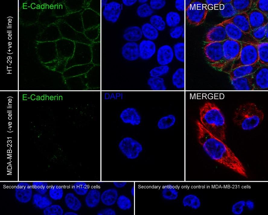
Immunocytochemistry analysis of HT-29 (positive) and MDA-MB-231 (negative) cells labeling E-Cadherin with E-Cadherin antibody at 1/2,000 dilution.
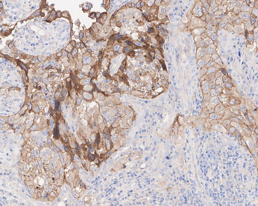
Immunohistochemical analysis of paraffin-embedded human lung carcinoma tissue with E-Cadherin antibody at 1/200 dilution.
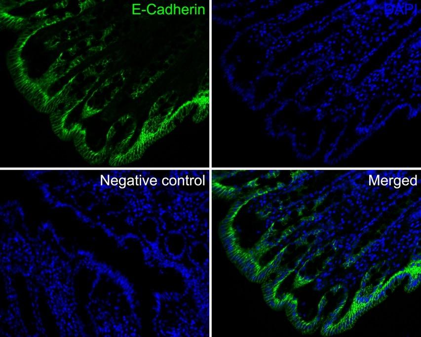
Application: IF-tissue;
Species: Human;
Site: Colon;
Sample: Paraffin-embedded section;
Antibody concentration: 1/200;
Species: Human;
Site: Colon;
Sample: Paraffin-embedded section;
Antibody concentration: 1/200;
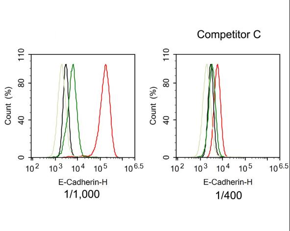
Flow cytometric analysis of HT-29 (positive, red) and MDA-MB-231 (negative, green) cells labeling E-Cadherin. Cells were fixed and permeabilized. Then stained with the primary antibody (red) at 1/1,000 dilution and competitor's antibody (red) at 1/400 dilution, compared with Rabbit IgG Isotype Control (HT-29 black, MDA-MB-231 light green). After incubation of the primary antibody at +4℃ for an hour, the cells were stained with a iFluor™ 488 conjugate-Goat anti-Rabbit IgG Secondary antibody at 1/1,000 dilution for 30 minutes at +4℃.
NOTE
Application
- WBWestern Blotting
- IHCImmunohistochemistry
- IFImmunofluorescence
- ICCImmunocytochemistry
- FCFlow Cytometry
- IPImmunoprecipitation
- EELISA
- DBDot Blotting
- ChIPChromatin Immunoprecipitation
- GICAGold Immunochromatography Assay
- NCNegative Control
Species Reactivity
- HuHuman
- MsMouse
- RtRat
- DmDrosophila melanogaster
- CCaenorhabditis elegans
- MkMonkey
- RbRabbit
- BBovine
- DDog
- PPig
- HmHamster
- ChHmChinese Hamster
- ChkChicken
- ShpSheep


