Product Detail
Product NameHDAC2 Rabbit mAb
Clone No.SY29-02
Host SpeciesRecombinant Rabbit
ClonalityMonoclonal
PurificationProA affinity purified
ApplicationsWB, ICC/IF, IHC, FC
Species ReactivityHu, Ms, Rt
Immunogen Descrecombinant protein
ConjugateUnconjugated
Other NamesD10Wsu179e antibody HD 2 antibody HD2 antibody HDAC 2 antibody Hdac2 antibody HDAC2_HUMAN antibody Histone deacetylase 2 (HD2) antibody Histone deacetylase 2 antibody OTTHUMP00000017046 antibody OTTHUMP00000227077 antibody OTTHUMP00000227078 antibody RPD3 antibody transcriptional regulator homolog RPD3 antibody YAF1 antibody YY1 associated factor 1 antibody YY1 transcription factor binding protein antibody Yy1bp antibody
Accession NoSwiss-Prot#:Q92769
Uniprot
Q92769
Gene ID
3066;
Calculated MW55 kDa
Sdspage MW55 kDa
Formulation1*TBS (pH7.4), 1%BSA, 40%Glycerol. Preservative: 0.05% Sodium Azide.
StorageStore at -20˚C
Application Details
WB: 1:1,000-1:2,000
IHC: 1:50-1:200
ICC: 1:50-1:200
FC: 1:50-1:100
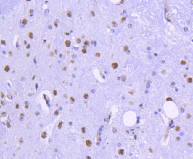
Immunohistochemical analysis of paraffin-embedded rat spinal cord tissue using anti-HDAC2 antibody. Counter stained with hematoxylin.
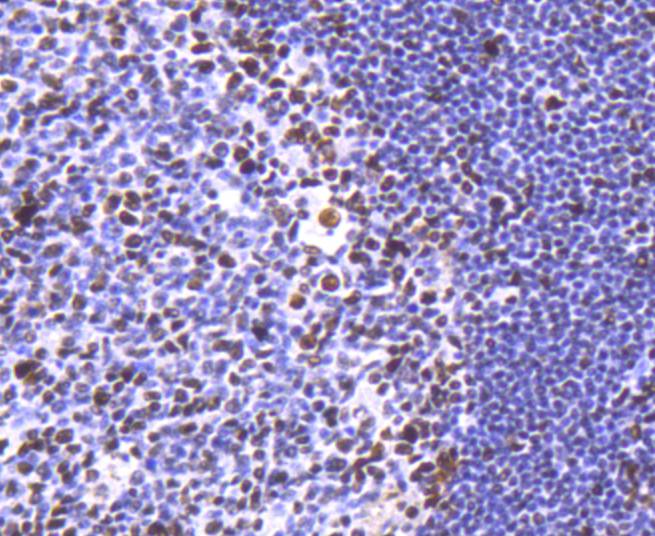
Immunohistochemical analysis of paraffin-embedded human tonsil tissue using anti-HDAC2 antibody. Counter stained with hematoxylin.
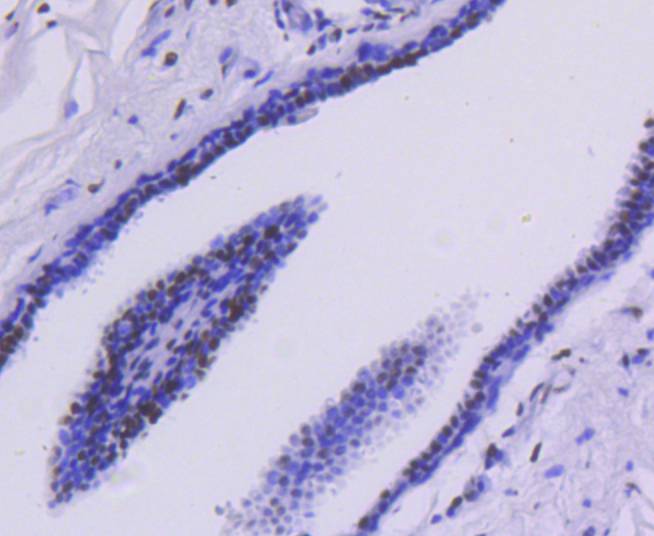
Immunohistochemical analysis of paraffin-embedded human breast carcinoma tissue using anti-HDAC2 antibody. Counter stained with hematoxylin.
Immunohistochemical analysis of paraffin-embedded mouse spinal cord tissue using anti-HDAC2 antibody. Counter stained with hematoxylin.
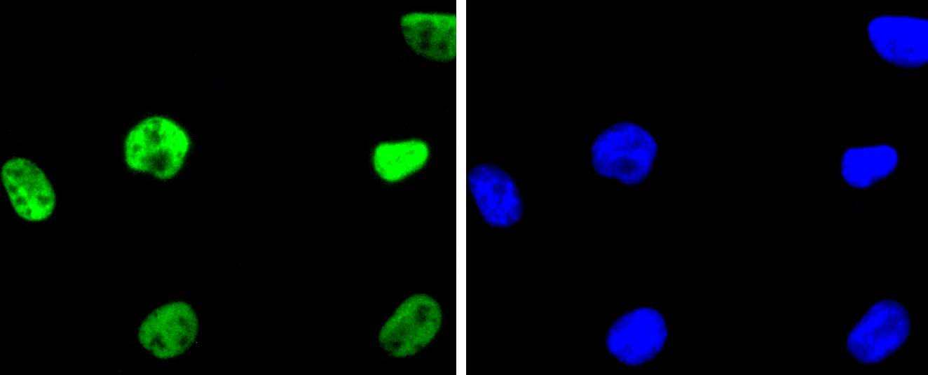
ICC staining HDAC2 in Hela cells (green). The nuclear counter stain is DAPI (blue). Cells were fixed in paraformaldehyde, permeabilised with 0.25% Triton X100/PBS.
ICC staining HDAC2 in SHG-44 cells (green). The nuclear counter stain is DAPI (blue). Cells were fixed in paraformaldehyde, permeabilised with 0.25% Triton X100/PBS.
ICC staining HDAC2 in 293 cells (green). The nuclear counter stain is DAPI (blue). Cells were fixed in paraformaldehyde, permeabilised with 0.25% Triton X100/PBS.
Flow cytometric analysis of Hela cells with HDAC2 antibody at 1/50 dilution (red) compared with an unlabelled control (cells without incubation with primary antibody; black). Alexa Fluor 488-conjugated goat anti rabbit IgG was used as the secondary antibody.
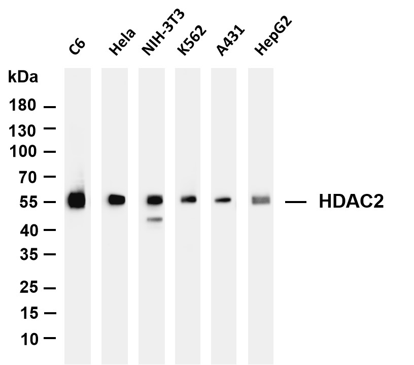
All lanes : HDAC2 Rabbit mAb at 1/1k dilution Lane 1 : C6 cell lysate Lane 2 : Hela cell lysate Lane 3 : NIH-3T3 cell lysate Lane 4 : K562 cell lysate Lane 5 : A431 cell lysate Lane 6 : HepG2 cell lysate Lysates/proteins at 20 µg per lane. Predicted band size: 55kDa Observed band size: 55kDa
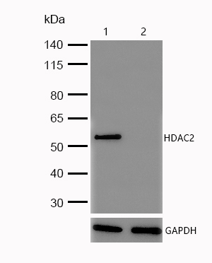
All lanes : HDAC2 Rabbit mAb at 1/1k dilution Lane 1 : Wild-type HAP1 cell lysate Lane 2 : HDAC2 knockout HAP1 cell lysate Lysates/proteins at 20 µg per lane.
In the intact cell, DNA closely associates with histones and other nuclear proteins to form chromatin. The remodeling of chromatin is believed to be a critical component of transcriptional regulation, and a major source of this remodeling is brought about by the acetylation of nucleosomal histones. Acetylation of lysine residues in the amino terminal tail domain of histone results in an allosteric change in the nucleosomal conformation and an increased accessibility to transcription factors by DNA. Conversely, the deacetylation of histones is associated with transcriptional silencing. Several mammalian proteins have been identified as nuclear histone acetylases, including GCN5, PCAF (for p300/CBP-associated factor), p300/CBP and the TFIID subunit TAF II p250. Mammalian HDAC1 (also designated HD1) and HDAC2 (also designated mammalian RPD3), both of which are related to the yeast transcriptional regulator Rpd3p, have been identified as histone deacetylases.
If you have published an article using product 48802, please notify us so that we can cite your literature.





 Yes
Yes


