Product Detail
Product NameCaMK Ⅱ Rabbit mAb
Clone No.SU03-57
Host SpeciesRecombinant Rabbit
Clonality Monoclonal
PurificationProA affinity purified
ApplicationsWB, ICC/IF, IHC
Species ReactivityHu, Ms, Rt
Immunogen Descrecombinant protein
ConjugateUnconjugated
Other NamesCalcium/calmodulin dependent protein kinase II alpha antibody
Calcium/calmodulin dependent protein kinase II beta antibody
Calcium/calmodulin dependent protein kinase II delta antibody
Calcium/calmodulin dependent protein kinase II gamma antibody
Calcium/calmodulin-dependent protein kinase type II subunit alpha antibody
CaM kinase II alpha antibody
CaM kinase II antibody
CaM kinase II beta antibody
CaM kinase II delta antibody
CaM kinase II gamma antibody
CaM kinase II subunit alpha antibody
CaMK-II subunit alpha antibody
CAMK2 antibody
Camk2a antibody
CAMK2B antibody
CAMK2D antibody
CAMK2G antibody
CAMKA antibody
KCC2A_HUMAN antibody
Accession NoSwiss-Prot#:Q13554
Uniprot
Q13554
Gene ID
816;
Calculated MW54 kDa
Formulation1*TBS (pH7.4), 1%BSA, 40%Glycerol. Preservative: 0.05% Sodium Azide.
StorageStore at -20˚C
Application Details
WB: 1:1,000-1:2,000
IHC: 1:50-1:200
ICC: 1:50-1:200
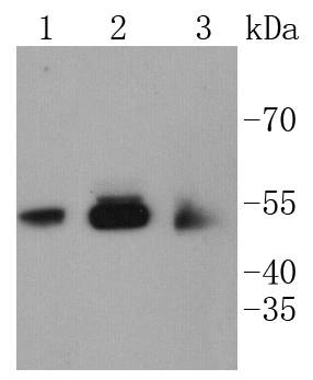
Western blot analysis of CaMK�� on different lysates using anti-CaMK�� antibody at 1/1,000 dilution. Positive control:
Lane 1: SH-SY-5Y
Lane 2: PC-12
Lane 3: SHG-44
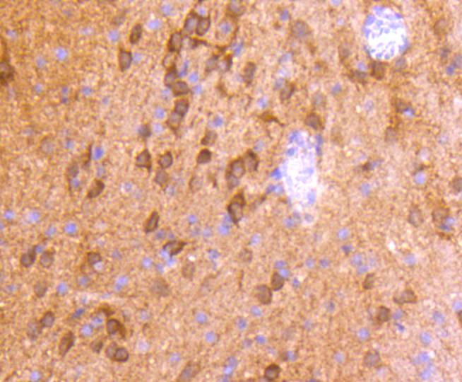
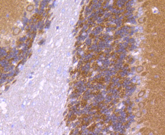
Immunohistochemical analysis of paraffin-embedded rat brain tissue using anti-CaMK�� antibody. Counter stained with hematoxylin.
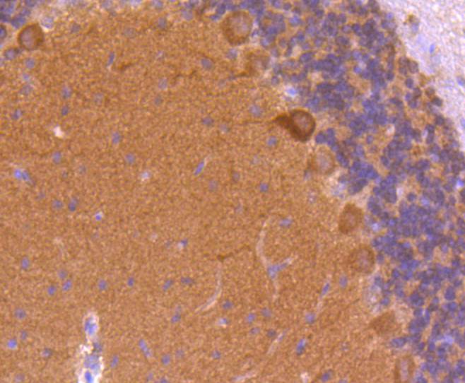
Immunohistochemical analysis of paraffin-embedded rat cerebellum tissue using anti-CaMK�� antibody. Counter stained with hematoxylin.
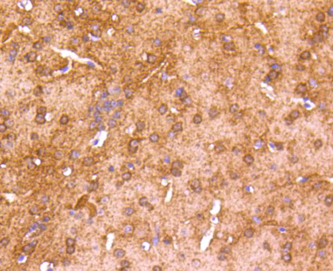
Immunohistochemical analysis of paraffin-embedded mouse brain tissue using anti-CaMK�� antibody. Counter stained with hematoxylin.
Immunohistochemical analysis of paraffin-embedded mouse cerebellum tissue using anti-CaMK�� antibody. Counter stained with hematoxylin.
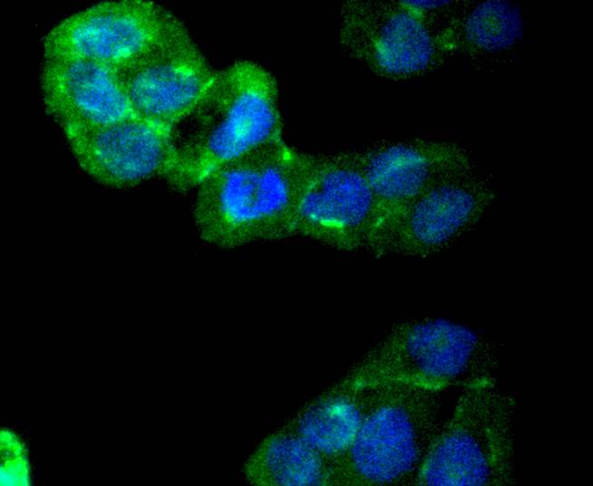
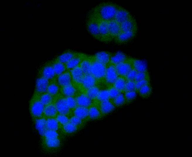
ICC staining CaMK�� in Hela cells (green). The nuclear counter stain is DAPI (blue). Cells were fixed in paraformaldehyde, permeabilised with 0.25% Triton X100/PBS.
ICC staining CaMK�� in PC-12 cells (green). The nuclear counter stain is DAPI (blue). Cells were fixed in paraformaldehyde, permeabilised with 0.25% Triton X100/PBS.
ICC staining CaMK�� in SHG-44 cells (green). The nuclear counter stain is DAPI (blue). Cells were fixed in paraformaldehyde, permeabilised with 0.25% Triton X100/PBS.
The Ca2+/calmodulin-dependent protein kinases (CaM kinases) comprise a structurally related subfamily of serine/threonine kinases which include CaMKI, CaMKII and CaMKIV. CaMKII is a ubiquitously expressed serine/threonine protein kinase that is activated by Ca2+and calmodulin (CaM) and has been implicated in regulation of the cell cycle and transcription. There are four CaMKII isozymes designated α, β, γ and δ, which may or may not be co-expressed in the same tissue type. CaMKIV is stimulated by Ca2+ and CaM but also requires phosphorylation by a CaMK for full activation. Stimulation of the T cell receptor CD3 signaling complex with an anti-CD3 monoclonal antibody leads to a 10-40 fold increase in CaMKIV activity. An additional kinase, CaMKK, functions to activate CaMKI through the specific phosphorylation of the regulatory Threonine residue at position 177.
If you have published an article using product 48831, please notify us so that we can cite your literature.



 Yes
Yes


