Product Detail
Product NameCathepsin D Rabbit mAb
Clone No.SU0360
Host SpeciesRecombinant Rabbit
Clonality Monoclonal
PurificationProA affinity purified
ApplicationsWB, ICC, IHC, IP, FC
Species ReactivityHu, Ms
Immunogen Descrecombinant protein
ConjugateUnconjugated
Other NamesCatD antibody CATD_HUMAN antibody Cathepsin D antibody Cathepsin D heavy chain antibody CD antibody Ceroid lipofuscinosis neuronal 10 antibody CLN10 antibody CPSD antibody ctsd antibody Epididymis secretory sperm binding protein Li 130P antibody HEL S 130P antibody Lysosomal aspartyl peptidase antibody Lysosomal aspartyl protease antibody MGC2311 antibody
Accession NoSwiss-Prot#:P07339
Uniprot
P07339
Gene ID
1509;
Calculated MW28/43/46 kDa
Formulation1*TBS (pH7.4), 1%BSA, 40%Glycerol. Preservative: 0.05% Sodium Azide.
StorageStore at -20˚C
Application Details
WB: 1:1,000-1:2,000
IHC: 1:100-1:500
ICC: 1:100-1:500
FC: 1:10-1:100
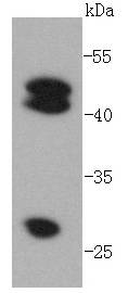
Western blot analysis of Cathepsin D on MCF-7 lysates using anti-Cathepsin D antibody at 1/1,000 dilution.
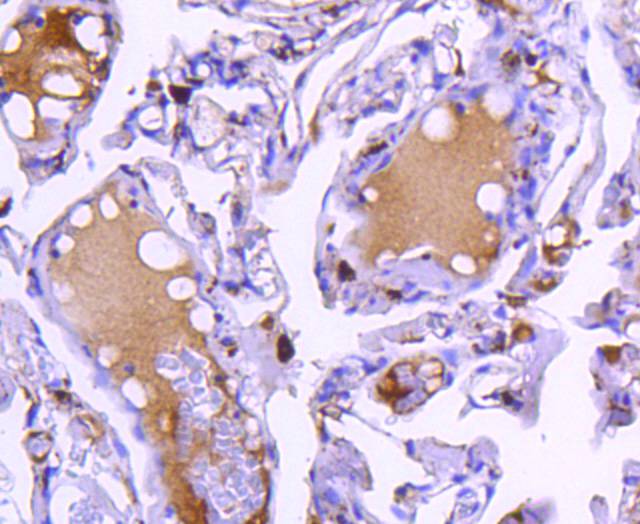
Immunohistochemical analysis of paraffin-embedded human lung tissue using anti-Cathepsin D antibody. Counter stained with hematoxylin.
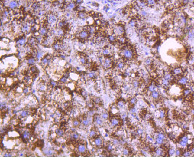
Immunohistochemical analysis of paraffin-embedded human liver tissue using anti-Cathepsin D antibody. Counter stained with hematoxylin.
Immunohistochemical analysis of paraffin-embedded human breast carcinoma tissue using anti-Cathepsin D antibody. Counter stained with hematoxylin.
Immunohistochemical analysis of paraffin-embedded human gastric cancer tissue using anti-Cathepsin D antibody. Counter stained with hematoxylin.
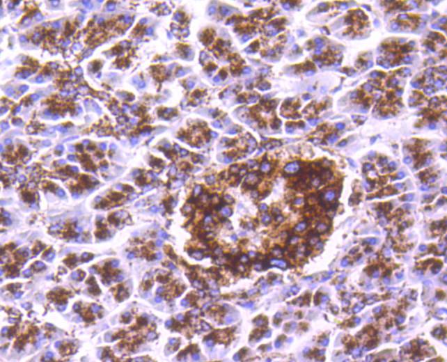
Immunohistochemical analysis of paraffin-embedded human pancreas tissue using anti-Cathepsin D antibody. Counter stained with hematoxylin.
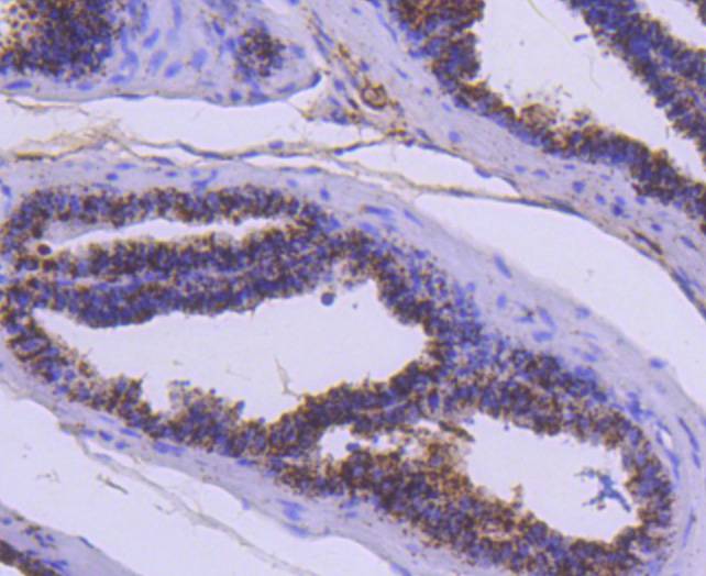
Immunohistochemical analysis of paraffin-embedded mouse prostate tissue using anti-Cathepsin D antibody. Counter stained with hematoxylin.
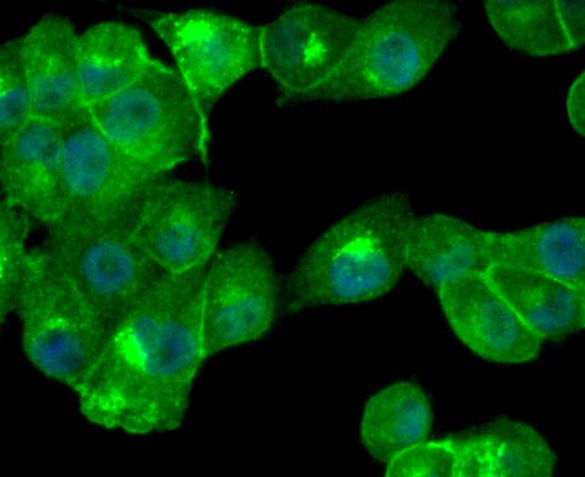
ICC staining Cathepsin D in PANC-1 cells (green). The nuclear counter stain is DAPI (blue). Cells were fixed in paraformaldehyde, permeabilised with 0.25% Triton X100/PBS.
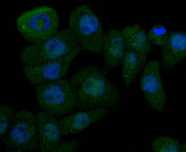
ICC staining Cathepsin D in AGS cells (green). The nuclear counter stain is DAPI (blue). Cells were fixed in paraformaldehyde, permeabilised with 0.25% Triton X100/PBS.
The cathepsin family of proteolytic enzymes contains several diverse classes of proteases. The cysteine protease class comprises cathepsins B, L, H, K, S, and O. The aspartyl protease class is composed of cathepsins D and E. Cathepsin G is in the serine protease class. Most cathepsins are lysosomal and each is involved in cellular metabolism, participating in various events such as peptide biosynthesis and protein degradation. Cathepsins may also cleave some protein precursors, thereby releasing regulatory peptides. The promoter region of the cathepsin D gene contains five Sp1 binding sites and four AP-2 binding sites.
If you have published an article using product 48833, please notify us so that we can cite your literature.




 Yes
Yes


