Product Detail
Product NameLactate Dehydrogenase Rabbit mAb
Clone No.SU39-06
ClonalityMonoclonal
PurificationProA affinity purified
ApplicationsWB, ICC/IF, IHC, IP, FC
Species ReactivityHu, Ms, Rt, zebrafish
Immunogen Descrecombinant protein
ConjugateUnconjugated
Other NamesCell proliferation-inducing gene 19 protein antibody
GSD11 antibody
L lactate dehydrogenase B chain antibody
L-lactate dehydrogenase A chain antibody
Lactate dehydrogenase A antibody
Lactate dehydrogenase B antibody
Lactate dehydrogenase H chain antibody
Lactate dehydrogenase M antibody
LDH A antibody
LDH B antibody
LDH H antibody
LDH heart subunit antibody
LDH M antibody
LDH muscle subunit antibody
LDH-A antibody
LDH-M antibody
LDH1 antibody
ldha antibody
LDHA_HUMAN antibody
LDHBD antibody
LDHM antibody
MS1111 antibody
PIG19 antibody
Proliferation inducing gene 19 antibody
Renal carcinoma antigen NY REN 46 antibody
Renal carcinoma antigen NY-REN-59 antibody
TRG 5 antibody
TRG5 antibody
Accession NoSwiss-Prot#:P00338
Uniprot
P00338
Gene ID
3939;
Calculated MW37 kDa
Concentration1mg/ml
Formulation1*TBS (pH7.4), 1%BSA, 40%Glycerol. Preservative: 0.05% Sodium Azide.
StorageStore at -20˚C
Application Details
WB: 1:1,000-5,000
IHC: 1:200-1:500
ICC: 1:50-1:200
FC: 1:50-1:100
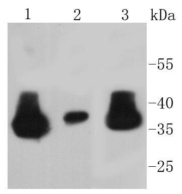
Western blot analysis of Lactate Dehydrogenase on different lysates using anti-Lactate Dehydrogenase antibody at 1/1,000 dilution. Positive control:
Lane 1: Hela
Lane 2: A549
Lane 3: MCF-7
Western blot analysis of Lactate Dehydrogenase on hybrid fish (crucian-carp) brain tissue lysate using anti-Lactate Dehydrogenase antibody at 1/500 dilution.
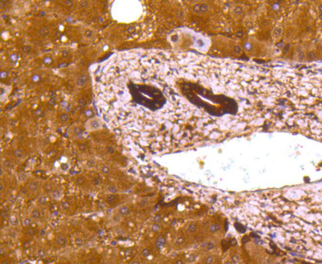
Immunohistochemical analysis of paraffin-embedded human liver tissue using anti-Lactate Dehydrogenase antibody. Counter stained with hematoxylin.
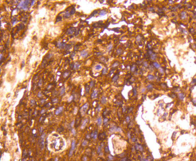
Immunohistochemical analysis of paraffin-embedded human breast carcinoma tissue using anti-Lactate Dehydrogenase antibody. Counter stained with hematoxylin.
Immunohistochemical analysis of paraffin-embedded mouse liver tissue using anti-Lactate Dehydrogenase antibody. Counter stained with hematoxylin.
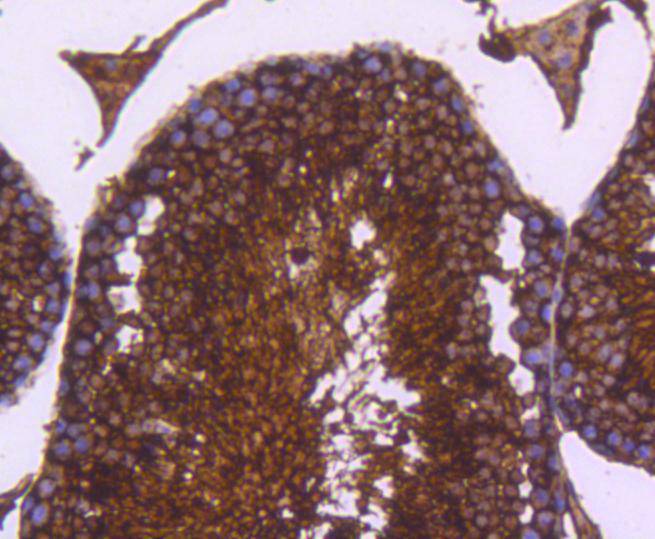
Immunohistochemical analysis of paraffin-embedded mouse testis tissue using anti-Lactate Dehydrogenase antibody. Counter stained with hematoxylin.
Immunohistochemical analysis of paraffin-embedded human liver cancer tissue using anti-Lactate Dehydrogenase antibody. Counter stained with hematoxylin.
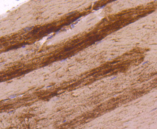
Immunohistochemical analysis of paraffin-embedded mouse skeletal muscle tissue using anti-Lactate Dehydrogenase antibody. Counter stained with hematoxylin.
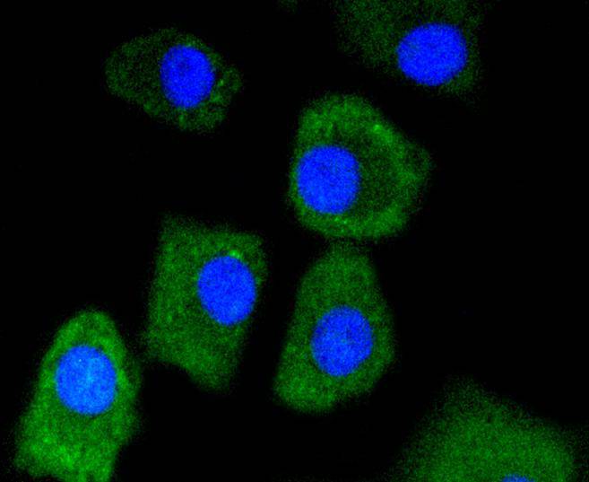
ICC staining Lactate Dehydrogenase in A549 cells (green). The nuclear counter stain is DAPI (blue). Cells were fixed in paraformaldehyde, permeabilised with 0.25% Triton X100/PBS.
ICC staining Lactate Dehydrogenase in A431 cells (green). The nuclear counter stain is DAPI (blue). Cells were fixed in paraformaldehyde, permeabilised with 0.25% Triton X100/PBS.
Flow cytometric analysis of Hela cells with Lactate Dehydrogenase antibody at 1/50 dilution (red) compared with an unlabelled control (cells without incubation with primary antibody; black).
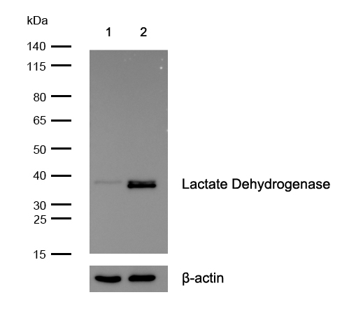
All lanes : Lactate Dehydrogenase Rabbit mAb at 1/1k dilution Lane 1 : Lactate Dehydrogenase knockout HAP1 cell lysate Lane 2 : Wild-type HAP1 cell lysate Lysates/proteins at 20 µg per lane.
The lactate dehydrogenase family (LDH) catalyzes the final step of anaerobic glycolysis, the conversion of L-lactate and NAD to pyruvate and NADH. The LDH family consists of three members, LDH-A, LDH-B and LDH-C, all of which form tetramers consisting four subunits. However, each family member displays a specific tissue distribution pattern with LDH-A and LDH-B predominant in several tissues, specifically LDH-A in muscle and LDH-B in heart, while LDH-C expression is confined to the testis and sperm. LDHs function as powerful markers for germ cell tumors. The genes encoding human LDH-A and LDH-C map to chromosome 11, while the human LDH-B gene maps to chromosome 12. Deficiency in the LDH-A gene is linked to exertional myoglobinuria.
If you have published an article using product 48839, please notify us so that we can cite your literature.







 Yes
Yes


