Product Detail
Product NameAurora A Rabbit mAb
Clone No.ST46-07
Host SpeciesRecombinant Rabbit
Clonality Monoclonal
PurificationProA affinity purified
ApplicationsWB, ICC/IF, FC
Species ReactivityHu, Ms
Immunogen Descrecombinant protein
ConjugateUnconjugated
Other NamesAIK antibody ARK-1 antibody ARK1 antibody AURA antibody Aurka antibody Aurora 2 antibody Aurora A antibody Aurora kinase A antibody Aurora-related kinase 1 antibody Aurora/IPL1 like kinase antibody Aurora/IPL1-related kinase 1 antibody AURORA2 antibody Breast tumor-amplified kinase antibody BTAK antibody hARK1 antibody IAK antibody IPL1 related kinase antibody MGC34538 antibody OTTHUMP00000031340 antibody OTTHUMP00000031341 antibody OTTHUMP00000031342 antibody OTTHUMP00000031343 antibody OTTHUMP00000031344 antibody OTTHUMP00000031345 antibody OTTHUMP00000166071 antibody OTTHUMP00000166072 antibody PPP1R47 antibody Protein phosphatase 1, regulatory subunit 47 antibody Serine/threonine kinase 15 antibody Serine/threonine kinase 6 antibody Serine/threonine-protein kinase 15 antibody Serine/threonine-protein kinase 6 antibody Serine/threonine-protein kinase aurora-A antibody STK15 antibody STK6 antibody STK6_HUMAN antibody STK7 antibody
Accession NoSwiss-Prot#:O14965
Uniprot
O14965
Gene ID
6790;
Calculated MW46 kDa
Formulation1*TBS (pH7.4), 1%BSA, 40%Glycerol. Preservative: 0.05% Sodium Azide.
StorageStore at -20˚C
Application Details
WB: 1:1,000-1:2,000
ICC: 1:100-1:500
FC: 1:50-1:100
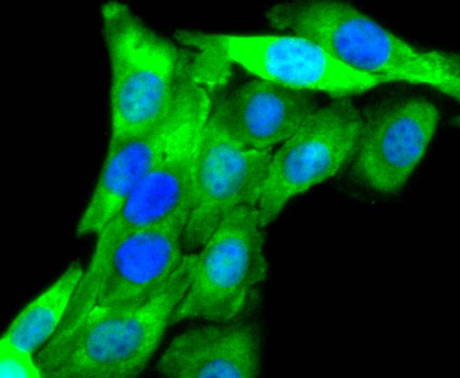
ICC staining Aurora A in SKOV-3 cells (green). The nuclear counter stain is DAPI (blue). Cells were fixed in paraformaldehyde, permeabilised with 0.25% Triton X100/PBS.
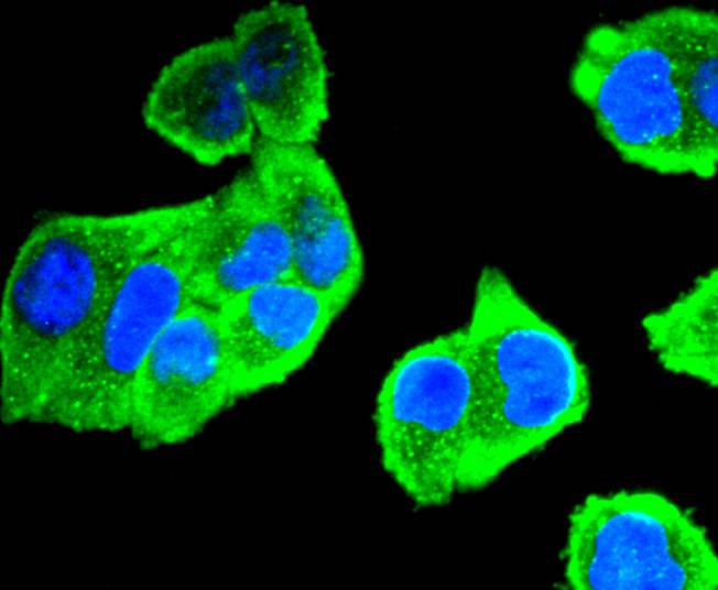
ICC staining Aurora A in Hela cells (green). The nuclear counter stain is DAPI (blue). Cells were fixed in paraformaldehyde, permeabilised with 0.25% Triton X100/PBS.
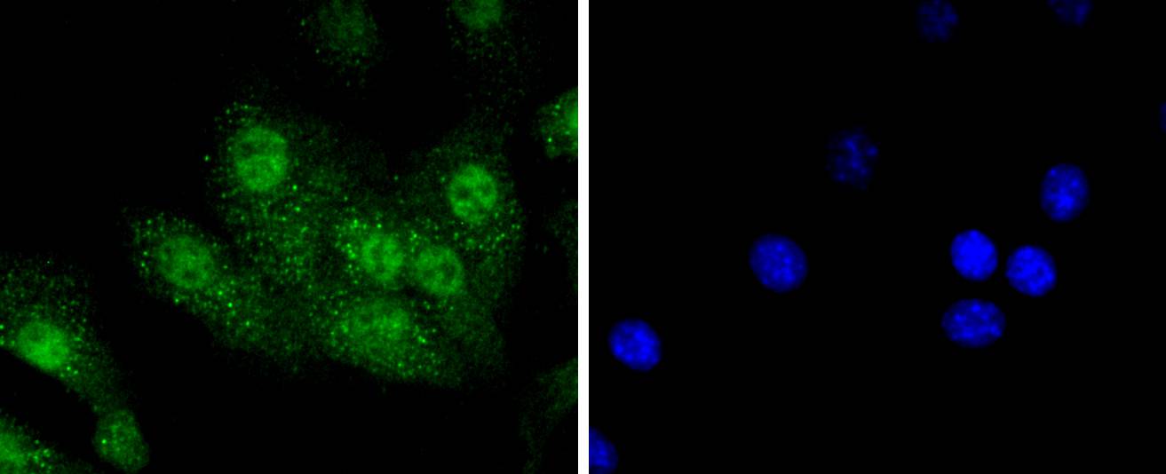
ICC staining Aurora A in NIH/3T3 cells (green). The nuclear counter stain is DAPI (blue). Cells were fixed in paraformaldehyde, permeabilised with 0.25% Triton X100/PBS.
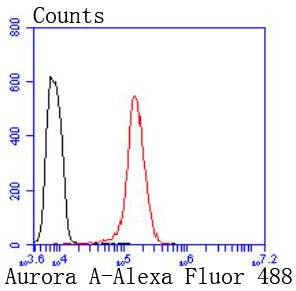
Flow cytometric analysis of Hela cells with Aurora A antibody at 1/50 dilution (red) compared with an unlabelled control (cells without incubation with primary antibody; black). Alexa Fluor 488-conjugated goat anti rabbit IgG was used as secondary antibody
Activation of the oncogenic protein kinase Aurora A regulates meiotic and mitotic cell cycles in eukaryotic cells. Specifically, Aurora A plays a key role in G2/M progression. Activation occurs via autophosphorylation, and while 14 sites are subject to this, only the threonine residue at position 295 is required for activity. Though autophosphorylation mediates activation, a number of other proteins influence activation, including the spindle assembly factor TPX2 and p53
If you have published an article using product 48861, please notify us so that we can cite your literature.


 Yes
Yes


