Location:
Home
>
Recombinant Rabbit Monoclonal Antibodies > Hexokinase 1 Rabbit mAb
Hexokinase 1 Rabbit mAb#48867

 Yes
Yes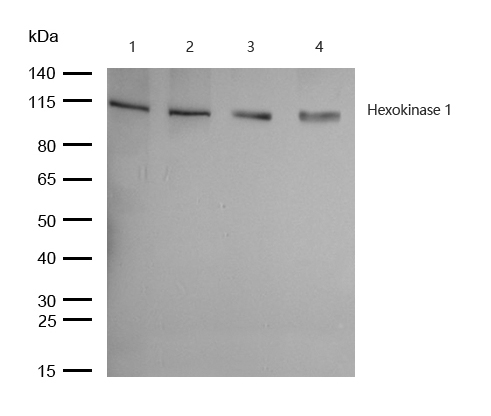
All lanes: Hexokinase 1 Rabbit mAb at 1/1k dilution
Lane 1 : 293 whole cell lysates
Lane 2 : SH-SY5Y whole cell lysatesLane 3 : Mouse heart lysatesLane 4 : Rat liver lysates
Lysates/proteins at 20 µg per lane.
Secondary
All lanes : Goat Anti-Rabbit IgG H&L (HRP) at 1/20000 dilution
Predicted band size: 102 kDa
Observed band size: 110 kDa
Exposure time: 13 seconds
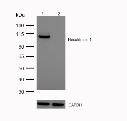
All lanes:Hexokinase 1 Rabbit mAb at 1/1k dilution
Lane 1 : Wild-type Hela cell lysate
Lane 2 : Hexokinase 1 knockdown Hela cell lysate
Lysates/proteins at 20 µg per lane.
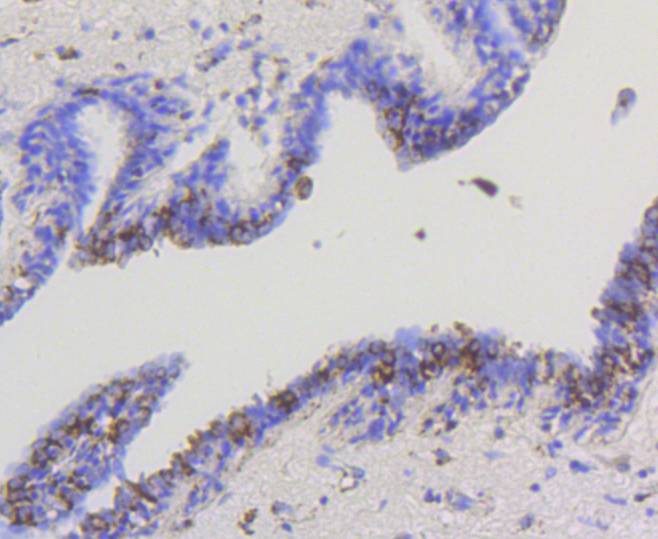
Formalin-fixed, paraffin-embedded human lung tissue stained for Hexokinase 1 using 48867 at 1/100 dilution in immunohistochemical analysis.
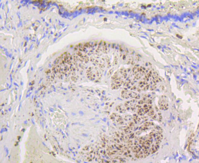
Formalin-fixed, paraffin-embedded human breast carcinoma tissue stained for Hexokinase 1 using 48867 at 1/100 dilution in immunohistochemical analysis.
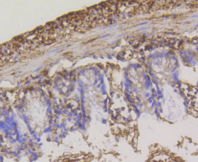
Formalin-fixed, paraffin-embedded mouse colon tissue stained for Hexokinase 1 using 48867 at 1/100 dilution in immunohistochemical analysis.
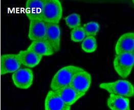
Immunocytochemistry/ Immunofluorescence Hexokinase 1 antibody (48867) ICC/IF staining of Hexokinase 1 in CRC cells. Cells were fixed with 4% Paraformaldehyde permeabilized with 0.1% Triton X-100. Samples were incubated with 48867 at a working dilution of 1/100. The secondary antibody was Alexa Fluor® 488 goat anti rabbit, used at a dilution of 1/500. Nuclei were counterstained with DAPI.

Immunocytochemistry/ Immunofluorescence Hexokinase 1 antibody (48867) ICC/IF staining of Hexokinase 1 in MCF-7 cells. Cells were fixed with 4% Paraformaldehyde permeabilized with 0.1% Triton X-100. Samples were incubated with 48867 at a working dilution of 1/100. The secondary antibody was Alexa Fluor® 488 goat anti rabbit, used at a dilution of 1/500. Nuclei were counterstained with DAPI.
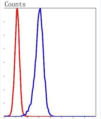
Flow Cytometry (Intracellular) Hexokinase 1 antibody (48867)
Flow cytometric analysis of K562 cells with Hexokinase 1 antibody at 1/50 dilution (blue) compared with an unlabelled control (cells without incubation with primary antibody,red). Alexa Fluor 488-conjugated goat anti rabbit IgG was used as the secondary antibody.
NOTE
Application
- WBWestern Blotting
- IHCImmunohistochemistry
- IFImmunofluorescence
- ICCImmunocytochemistry
- FCFlow Cytometry
- IPImmunoprecipitation
- EELISA
- DBDot Blotting
- ChIPChromatin Immunoprecipitation
- GICAGold Immunochromatography Assay
- NCNegative Control
Species Reactivity
- HuHuman
- MsMouse
- RtRat
- DmDrosophila melanogaster
- CCaenorhabditis elegans
- MkMonkey
- RbRabbit
- BBovine
- DDog
- PPig
- HmHamster
- ChHmChinese Hamster
- ChkChicken
- ShpSheep


