Location:
Home
>
Recombinant Rabbit Monoclonal Antibodies > ATP citrate lyase Rabbit mAb
ATP citrate lyase Rabbit mAb#48874

 Yes
Yes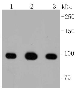
Western blot analysis of ATP citrate lyase on different lysates using anti-ATP citrate lyase antibody at 1/1,000 dilution. Positive control: Lane 1: Mouse kidney Lane 2: Mouse colon Lane 3: MCF-7

Immunohistochemical analysis of paraffin-embedded human thyroid tissue using anti-ATP citrate lyase citrate lyase antibody. Counter stained with hematoxylin.

Immunohistochemical analysis of paraffin-embedded human breast carcinoma tissue using anti-ATP citrate lyase antibody. Counter stained with hematoxylin.

Immunohistochemical analysis of paraffin-embedded mouse thyroid tissue using anti-ATP citrate lyase antibody. Counter stained with hematoxylin.

Immunohistochemical analysis of paraffin-embedded mouse kidney tissue using anti-ATP citrate lyase antibody. Counter stained with hematoxylin.
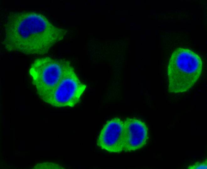
ICC staining ATP citrate lyase in Hela cells (green). The nuclear counter stain is DAPI (blue). Cells were fixed in paraformaldehyde, permeabilised with 0.25% Triton X100/PBS.

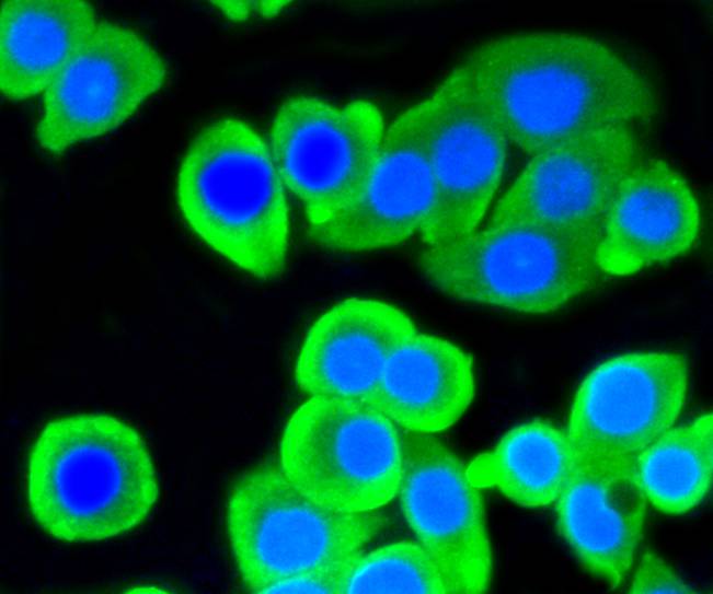
ICC staining ATP citrate lyase in A549 cells (green). The nuclear counter stain is DAPI (blue). Cells were fixed in paraformaldehyde, permeabilised with 0.25% Triton X100/PBS.
ICC staining ATP citrate lyase in CRC cells (green). The nuclear counter stain is DAPI (blue). Cells were fixed in paraformaldehyde, permeabilised with 0.25% Triton X100/PBS.
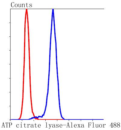
Flow cytometric analysis of Hela cells with ATP citrate lyase antibody at 1/50 dilution (blue) compared with an unlabelled control (cells without incubation with primary antibody; red). Alexa Fluor 488-conjugated goat anti rabbit IgG was used as the secondary antibody.
NOTE
Application
- WBWestern Blotting
- IHCImmunohistochemistry
- IFImmunofluorescence
- ICCImmunocytochemistry
- FCFlow Cytometry
- IPImmunoprecipitation
- EELISA
- DBDot Blotting
- ChIPChromatin Immunoprecipitation
- GICAGold Immunochromatography Assay
- NCNegative Control
Species Reactivity
- HuHuman
- MsMouse
- RtRat
- DmDrosophila melanogaster
- CCaenorhabditis elegans
- MkMonkey
- RbRabbit
- BBovine
- DDog
- PPig
- HmHamster
- ChHmChinese Hamster
- ChkChicken
- ShpSheep


