Welcome to Sabbiotech!     
Email:info@signalwayantibody.com
Location: Home > Recombinant Rabbit Monoclonal Antibodies > Chk1 Rabbit mAb

Chk1 Rabbit mAb#48908

Availability: Yes
Total Amount:$
Product Detail

Product NameChk1 Rabbit mAb

Clone No.ST57-09

Host SpeciesRecombinant Rabbit

Clonality Monoclonal

PurificationProA affinity purified

ApplicationsWB, ICC/IF, IHC, FC

Species ReactivityHu

Immunogen Descrecombinant protein

ConjugateUnconjugated

Other NamesC85740 antibody
Cell cycle checkpoint kinase antibody
Checkpoint , S. pombe, homolog of, 1 antibody
Checkpoint kinase 1 antibody
Checkpoint kinase 1 homolog (S. pombe) antibody
CHEK 1 antibody
Chek1 antibody
Chk 1 antibody
Chk1 antibody
CHK1 checkpoint homolog (S. pombe) antibody
CHK1_HUMAN antibody
EC 2.7.11.1 antibody
rad27 antibody
Serine/threonine protein kinase Chk1 antibody
Serine/threonine-protein kinase CHK1 antibody
STT3, subunit of the oligosaccharyltransferase complex, homolog A (S. cerevisiae) antibody

Accession NoSwiss-Prot#:O14757

Uniprot O14757

Gene ID 1111;

Calculated MW54 kDa

Formulation1*TBS (pH7.4), 1%BSA, 40%Glycerol. Preservative: 0.05% Sodium Azide.

StorageStore at -20˚C

Application Details
WB: 1:1,000-1:2,000
IHC: 1:50-1:200
ICC: 1:50-1:200

FC: 1:50-1:100
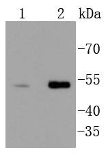
Western blot analysis of Chk1 on different lysates using anti-Chk1 antibody at 1/1,000 dilution. Positive control:
Lane 1: Hela
Lane 2: PC-3M
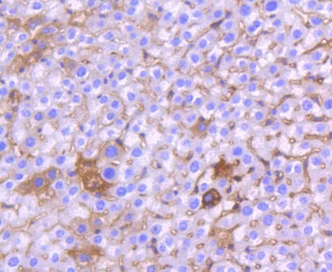
Immunohistochemical analysis of paraffin-embedded mouse liver tissue using anti-Chk1 antibody. Counter stained with hematoxylin.
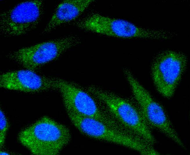
ICC staining Chk1 in Hela cells (green). The nuclear counter stain is DAPI (blue). Cells were fixed in paraformaldehyde, permeabilised with 0.25% Triton X100/PBS.
ICC staining Chk1 in MCF-7 cells (green). The nuclear counter stain is DAPI (blue). Cells were fixed in paraformaldehyde, permeabilised with 0.25% Triton X100/PBS.
ICC staining Chk1 in PC-3M cells (green). The nuclear counter stain is DAPI (blue). Cells were fixed in paraformaldehyde, permeabilised with 0.25% Triton X100/PBS.
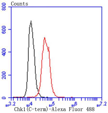
Flow cytometric analysis of Hela cells with Chk1 antibody at 1/50 dilution (red) compared with an unlabelled control (cells without incubation with primary antibody; black). Alexa Fluor 488-conjugated goat anti rabbit IgG was used as the secondary antibody
Cell cycle events are regulated by the sequential activation and deactivation of cyclin dependent kinases (Cdks) and by proteolysis of cyclins. Chk1 and Chk2 are involved in these processes as regulators of Cdks. Chk1 and Chk2 both function as essential components in the G2 DNA damage checkpoint by phosphorylating Cdc25C in response to DNA damage. Phosphorylation inhibits Cdc25C activity, thereby blocking mitosis. Cdc25A, Cdc25B and Cdc25C protein tyrosine phosphatases function as mitotic activators by dephosphorylating Cdc2 p34 on regulatory tyrosine residues. It has also been shown that Chk1 can phosphorylate Wee1 in vitro, providing evidence that the hyperphosphorylated form of Wee1, seen in cells delayed by Chk1 overexpression, is due to phosphorylation by Chk1.

If you have published an article using product 48908, please notify us so that we can cite your literature.

NOTE

Application

  • WBWestern Blotting
  • IHCImmunohistochemistry
  • IFImmunofluorescence
  • ICCImmunocytochemistry
  • FCFlow Cytometry
  • IPImmunoprecipitation
  • EELISA
  • DBDot Blotting
  • ChIPChromatin Immunoprecipitation
  • GICAGold Immunochromatography Assay
  • NCNegative Control

Species Reactivity

  • HuHuman
  • MsMouse
  • RtRat
  • DmDrosophila melanogaster
  • CCaenorhabditis elegans
  • MkMonkey
  • RbRabbit
  • BBovine
  • DDog
  • PPig
  • HmHamster
  • ChHmChinese Hamster
  • ChkChicken
  • ShpSheep
;