Product Detail
Product NameVAV2 Rabbit mAb
Clone No.SC68-03
Host SpeciesRecombinant Rabbit
Clonality Monoclonal
PurificationProA affinity purified
ApplicationsWB, ICC, IHC, IP
Species ReactivityHu, Ms, Rt
Immunogen Descrecombinant protein
ConjugateUnconjugated
Other NamesGuanine nucleotide exchange factor VAV2 antibody Oncogene VAV2 antibody Protein vav 2 antibody VAV 2 antibody Vav 2 oncogene antibody VAV-2 antibody VAV2 antibody VAV2 oncogene antibody VAV2_HUMAN antibody
Accession NoSwiss-Prot#:P52735
Uniprot
P52735
Gene ID
7410;
Calculated MW100 kDa
Formulation1*TBS (pH7.4), 1%BSA, 40%Glycerol. Preservative: 0.05% Sodium Azide.
StorageStore at -20˚C
Application Details
WB: 1:1,000-5,000
IHC: 1:50-1:200
ICC: 1:50-1:200
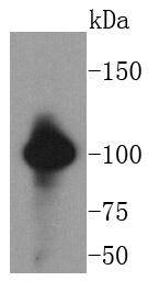
Western blot analysis of VAV2 on SW480 cells lysates using anti-VAV2 antibody at 1/1,000 dilution.
Immunohistochemical analysis of paraffin-embedded rat brain tissue using anti-VAV2 antibody. Counter stained with hematoxylin.
Immunohistochemical analysis of paraffin-embedded mouse colon tissue using anti-VAV2 antibody. Counter stained with hematoxylin.
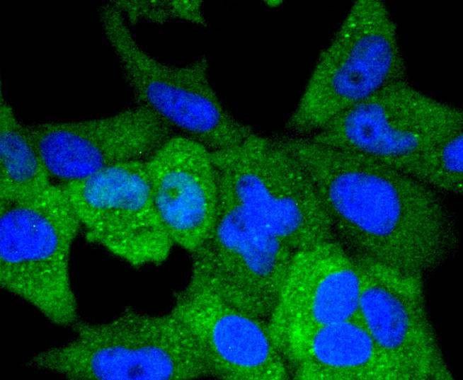
ICC staining VAV2 in Hela cells (green). The nuclear counter stain is DAPI (blue). Cells were fixed in paraformaldehyde, permeabilised with 0.25% Triton X100/PBS.
ICC staining VAV2 in NIH/3T3 cells (green). The nuclear counter stain is DAPI (blue). Cells were fixed in paraformaldehyde, permeabilised with 0.25% Triton X100/PBS.
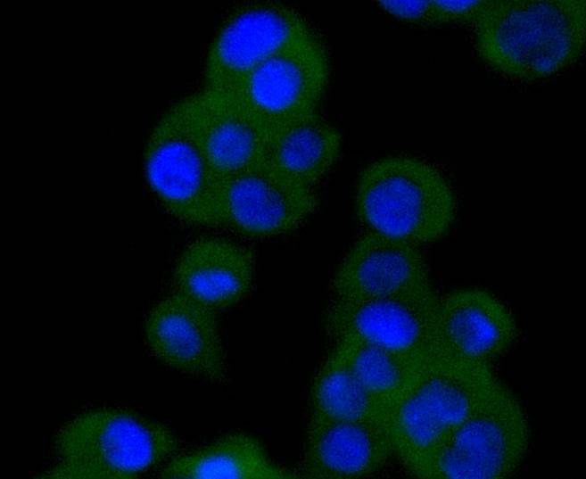
ICC staining VAV2 in SW480 cells (green). The nuclear counter stain is DAPI (blue). Cells were fixed in paraformaldehyde, permeabilised with 0.25% Triton X100/PBS.
The Vav gene was originally identified on the basis of its oncogenic activation during the course of gene transfer assays. The major translational product of the Vav proto-oncogene has been identified as a protein containing an array of structural motifs. This protein, known as Vav, Vav1 or p95Vav, contains an N-terminal helix-loop-helix domain and a leucine zipper motif similar to that of Myc family proteins that, if deleted, causes oncogenic activation. In addition, Vav contains an SH2 domain, which could indicate its role as a substrate for tyrosine kinases. Expression of Vav is limited exclusively to cells of hematopoietic origin, including those of the erythroid, lymphoid and myeloid lineages. These results suggest that Vav may represent a new type of signal transduction molecule involved in the transduction of tyrosine phosphorylation signaling into transcriptional events. Vav2 is a member of the Vav family of oncoproteins and acts as a guanosine nucleotide exchange factor (GEF) for RhoG and RhoA-like GTPases in a phosphotyrosine-dependent manner.
If you have published an article using product 48996, please notify us so that we can cite your literature.


 Yes
Yes


