Product Detail
Product NameCalnexin Rabbit mAb
Clone No.SN20-54
Host SpeciesRecombinant Rabbit
Clonality Monoclonal
PurificationProA affinity purified
ApplicationsWB, ICC/IF, IHC, FC
Species ReactivityHu, Rt
Immunogen Descrecombinant protein
ConjugateUnconjugated
Other NamesCalnexin antibody CALX_HUMAN antibody CANX antibody CNX antibody FLJ26570 antibody Histocompatibility complex class I antigen binding protein p88 antibody IP90 antibody Major histocompatibility complex class I antigen-binding protein p88 antibody p90 antibody
Accession NoSwiss-Prot#:P27824
Uniprot
P27824
Gene ID
821;
Calculated MW90 kDa
Formulation1*TBS (pH7.4), 1%BSA, 40%Glycerol. Preservative: 0.05% Sodium Azide.
StorageStore at -20˚C
Application Details
WB: 1:1,000-5,000
IHC: 1:50-1:200
ICC: 1:100-1:500
FC: 1:50-1:100
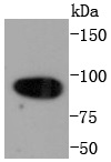
Western blot analysis of Calnexin on Hela cells lysates using anti-Calnexin antibody at 1/1,000 dilution.
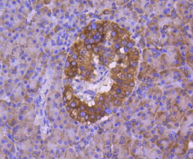
Immunohistochemical analysis of paraffin-embedded human pancreas tissue using anti-Calnexin antibody. Counter stained with hematoxylin.
Immunohistochemical analysis of paraffin-embedded rat heart tissue using anti-Calnexin antibody. Counter stained with hematoxylin.
Immunohistochemical analysis of paraffin-embedded rat pancreas tissue using anti-Calnexin antibody. Counter stained with hematoxylin.
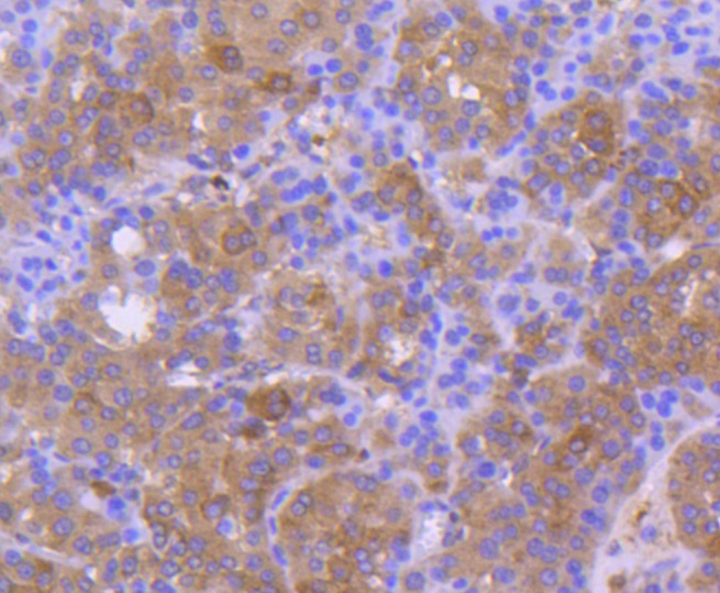
Immunohistochemical analysis of paraffin-embedded human liver cancer tissue using anti-Calnexin antibody. Counter stained with hematoxylin.
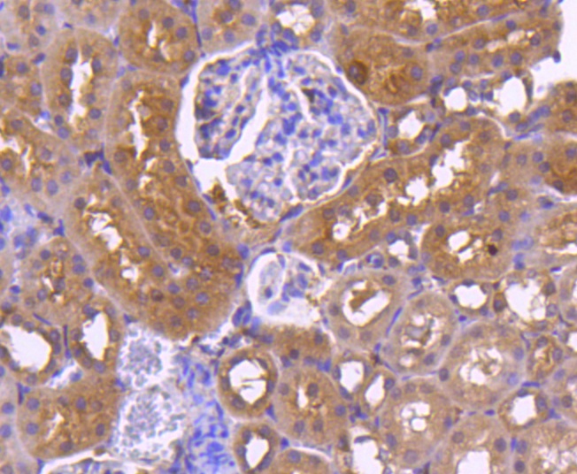
Immunohistochemical analysis of paraffin-embedded rat kidney tissue using anti-Calnexin antibody. Counter stained with hematoxylin.
Immunohistochemical analysis of paraffin-embedded human kidney tissue using anti-Calnexin antibody. Counter stained with hematoxylin.
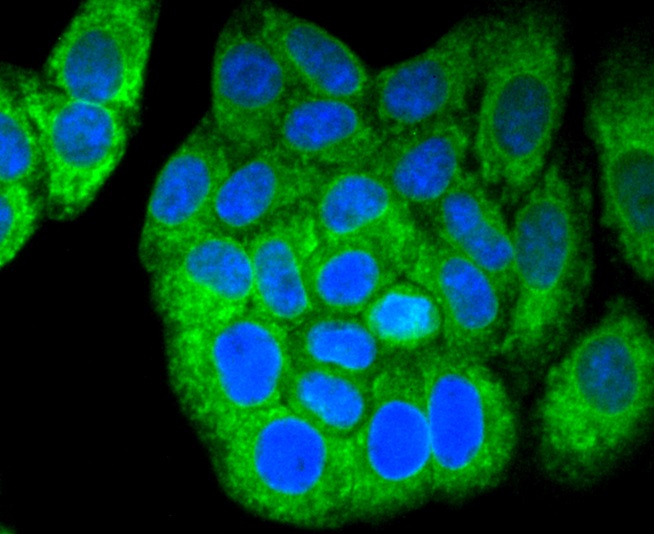
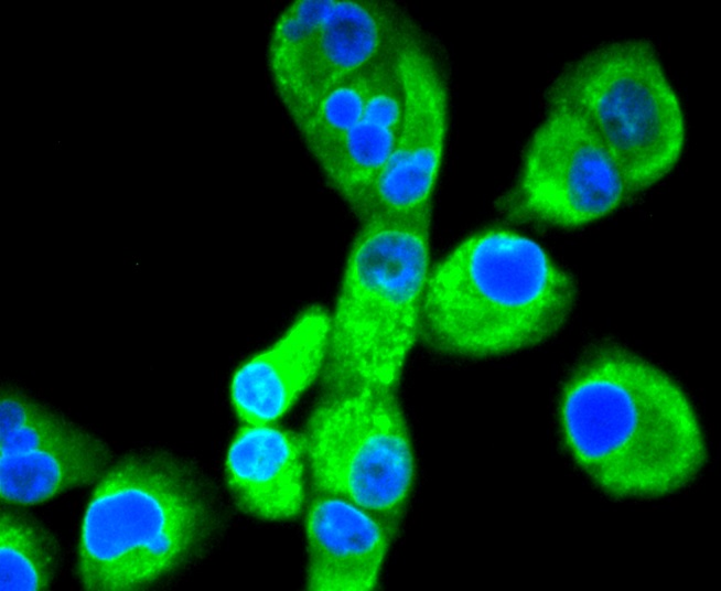
ICC staining Calnexin in Hela cells (green). The nuclear counter stain is DAPI (blue). Cells were fixed in paraformaldehyde, permeabilised with 0.25% Triton X100/PBS.
ICC staining Calnexin in HepG2 cells (green). The nuclear counter stain is DAPI (blue). Cells were fixed in paraformaldehyde, permeabilised with 0.25% Triton X100/PBS.
ICC staining Calnexin in PANC-1 cells (green). The nuclear counter stain is DAPI (blue). Cells were fixed in paraformaldehyde, permeabilised with 0.25% Triton X100/PBS.
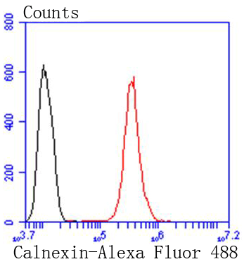
Flow cytometric analysis of Hela cells with Calnexin antibody at 1/50 dilution (red) compared with an unlabelled control (cells without incubation with primary antibody; black). Alexa Fluor 488-conjugated goat anti rabbit IgG was used as the secondary antibody
Calnexin and Calregulin (also called calreticulin) are calcium-binding proteins that are localized to the endoplasmic reticulum, Calnexin to the membrane and Calregulin to the lumen. Calnexin is a type I membrane protein that interacts with newly synthesized glycoproteins in the endoplasmic reticulum. It may play a role in assisting with protein assembly and in retaining unassembled protein subunits in the endoplasmic reticulum. Calregulin has both low- and high-affinity calcium-binding sites. Neither Calnexin nor Calregulin contains the calcium-binding E-F hand motif found in calmodulins. Calnexin and Calregulin are important for the maturation of glycoproteins in the endoplasmic reticulum and appear to bind many of the same proteins.
If you have published an article using product 49102, please notify us so that we can cite your literature.






 Yes
Yes


