Product Detail
Product NameIRF3 Rabbit mAb
Clone No.SD2062
Host SpeciesRecombinant Rabbit
Clonality Monoclonal
PurificationProA affinity purified
ApplicationsWB, ICC/IF, IHC, FC
Species ReactivityHu, Ms, Rt
Immunogen Descrecombinant protein
ConjugateUnconjugated
Other NamesIIAE7 antibody
Interferon regulatory factor 3 antibody
IRF 3 antibody
IRF-3 antibody
IRF3 antibody
IRF3_HUMAN antibody
MGC94729 antibody
Accession NoSwiss-Prot#:Q14653
Uniprot
Q14653
Gene ID
3661;
Calculated MW47 kDa
Formulation1*TBS (pH7.4), 1%BSA, 40%Glycerol. Preservative: 0.05% Sodium Azide.
StorageStore at -20˚C
Application Details
WB: 1:1,000-5,000
IHC: 1:50-1:200
ICC: 1:50-1:200
FC: 1:50-1:100
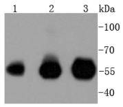
Western blot analysis of IRF3 on different lysates using anti-IRF3 antibody at 1/1,000 dilution. Positive control:
Lane 1: Hela
Lane 2: Jurkat
Lane 3: THP-1
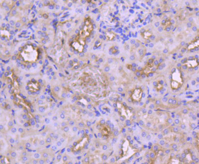
Immunohistochemical analysis of paraffin-embedded mouse kidney tissue using anti-IRF3 antibody. Counter stained with hematoxylin.
Immunohistochemical analysis of paraffin-embedded mouse pancreas tissue using anti-IRF3 antibody. Counter stained with hematoxylin.
Immunohistochemical analysis of paraffin-embedded human breast carcinoma tissue using anti-IRF3 antibody. Counter stained with hematoxylin.
Immunohistochemical analysis of paraffin-embedded human kidney tissue using anti-IRF3 antibody. Counter stained with hematoxylin.
Immunohistochemical analysis of paraffin-embedded human pancreas tissue using anti-IRF3 antibody. Counter stained with hematoxylin.
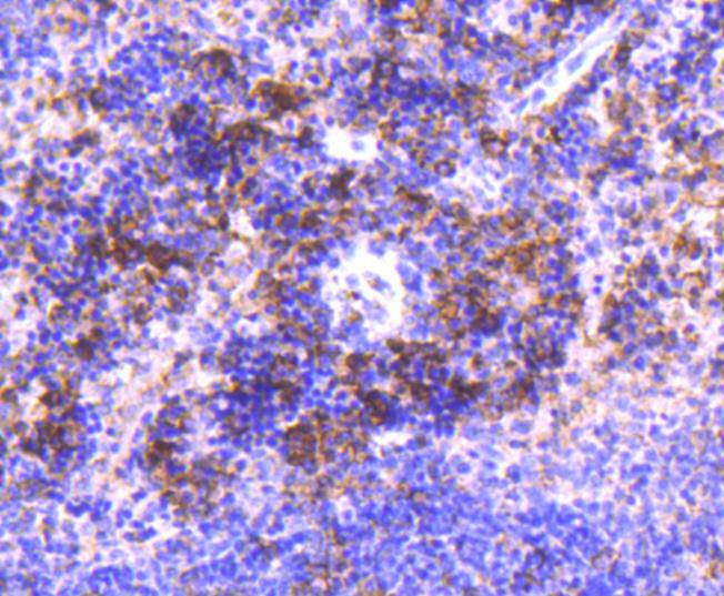
Immunohistochemical analysis of paraffin-embedded human tonsil tissue using anti-IRF3 antibody. Counter stained with hematoxylin.
Immunohistochemical analysis of paraffin-embedded mouse spleen tissue using anti-IRF3 antibody. Counter stained with hematoxylin.
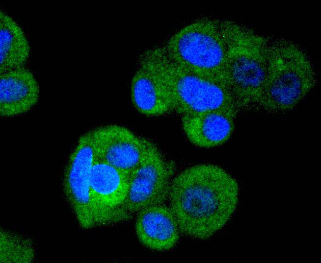
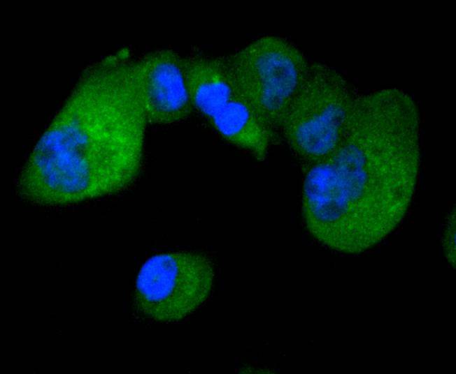
ICC staining IRF3 in Hela cells (green). The nuclear counter stain is DAPI (blue). Cells were fixed in paraformaldehyde, permeabilised with 0.25% Triton X100/PBS.
ICC staining IRF3 in MCF-7 cells (green). The nuclear counter stain is DAPI (blue). Cells were fixed in paraformaldehyde, permeabilised with 0.25% Triton X100/PBS.
ICC staining IRF3 in PANC-1 cells (green). The nuclear counter stain is DAPI (blue). Cells were fixed in paraformaldehyde, permeabilised with 0.25% Triton X100/PBS.
Interferon regulatory factor-1 (IRF-1) and IRF-2 have been identified as novel DNA-binding factors that function as regulators of both type I interferon (interferon-α and β) and interferon-inducible genes. The two factors are structurally related, particularly in their N-terminal regions, which confer DNA binding specificity. In addition, both bind to the same sequence within the promoters of interferon-α and interferon-β genes. IRF-1 functions as an activator of interferon transcription, while IRF-2 binds to the same cis elements and represses IRF-1 action. IRF-1 and IRF-2 have been reported to act in a mutually antagonistic manner in regulating cell growth; overexpression of the repressor IRF-2 leads to cell transformation while concomitant overexpression of IRF-1 causes reversion. IRF-1 and IRF-2 are members of a larger family of DNA binding proteins that includes IRF-3, IRF-4, IRF-5, IRF-6, IRF-7, ISGF-3γ p48 and IFN consensus sequence-binding protein (ICSBP).
If you have published an article using product 49122, please notify us so that we can cite your literature.





 Yes
Yes


