Welcome to Sabbiotech!     
Email:info@signalwayantibody.com
Location: Home > Recombinant Rabbit Monoclonal Antibodies > ERK5 Rabbit mAb

ERK5 Rabbit mAb#49183

Availability: Yes
Total Amount:$
Product Detail

Product NameERK5 Rabbit mAb

Clone No.SD2084

Host SpeciesRecombinant Rabbit

Clonality Monoclonal

PurificationProA affinity purified

ApplicationsWB, ICC/IF, IP, FC

Species ReactivityHu, Ms, Rt

Immunogen Descrecombinant protein

ConjugateUnconjugated

Other NamesBig MAP kinase 1 antibody
BMK 1 antibody
BMK 1 kinase antibody
BMK-1 antibody
BMK1 antibody
BMK1 Kinase antibody
EC 2.7.11.24 antibody
ERK 4 antibody
ERK 5 antibody
ERK-5 antibody
ERK4 antibody
ERK5 antibody
Extracellular signal regulated kinase 5 antibody
Extracellular signal-regulated kinase 5 antibody
MAP kinase 7 antibody
MAPK 7 antibody
MAPK7 antibody
Mitogen activated protein kinase 7 antibody
Mitogen-activated protein kinase 7 antibody
MK07_HUMAN antibody
OTTHUMP00000065906 antibody
OTTHUMP00000065907 antibody
PRKM 7 antibody
PRKM7 antibody
PROTEIN KINASE, MITOGEN-ACTIVATED, 7 antibody

Accession NoSwiss-Prot#:Q13164

Uniprot Q13164

Gene ID 5598;

Calculated MW115 kDa

Formulation1*TBS (pH7.4), 1%BSA, 40%Glycerol. Preservative: 0.05% Sodium Azide.

StorageStore at -20˚C

Application Details
WB: 1:1,000-1:2,000

ICC: 1:100-1:500

FC: 1:50-1:100
Western blot analysis of ERK5 on Hela cells lysates using anti-ERK5 antibody at 1/1,000 dilution.
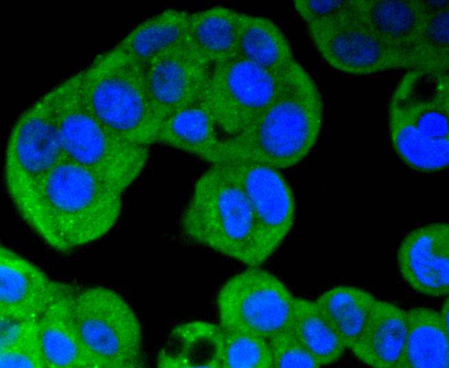
ICC staining ERK5 in Hela cells (green). The nuclear counter stain is DAPI (blue). Cells were fixed in paraformaldehyde, permeabilised with 0.25% Triton X100/PBS.
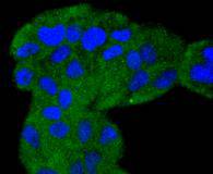
ICC staining ERK5 in MCF-7 cells (green). The nuclear counter stain is DAPI (blue). Cells were fixed in paraformaldehyde, permeabilised with 0.25% Triton X100/PBS.
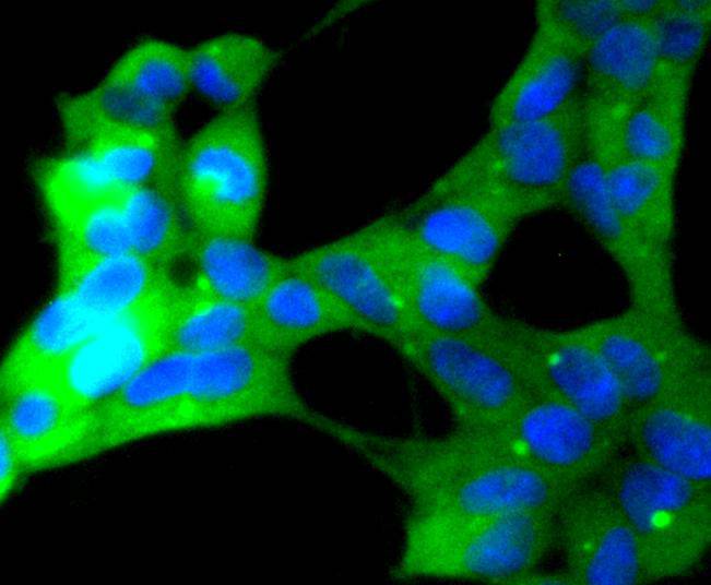
ICC staining ERK5 in 293 cells (green). The nuclear counter stain is DAPI (blue). Cells were fixed in paraformaldehyde, permeabilised with 0.25% Triton X100/PBS.
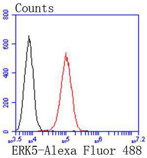
Flow cytometric analysis of A549 cells with ERK5 antibody at 1/50 dilution (red) compared with an unlabelled control (cells without incubation with primary antibody; black). Alexa Fluor 488-conjugated goat anti rabbit IgG was used as the secondary antibody.
The activation of signal transduction pathways by growth factors, hormones and neurotransmitters is mediated through two closely related MAP kinases, p44 and p42, designated extracellular-signal related kinase 1 (ERK 1) and ERK 2, respectively. ERK proteins are regulated by dual phosphorylation at specific tyrosine and threonine sites mapping within a characteristic Thr-Glu-Tyr motif. Phosphorylation at both the Thr and Tyr residues is required for full enzymatic activation. In response to activation, MAP kinases phosphorylate downstream components on serine and threonine. Upstream MAP kinase regulators include MAP kinase kinase (MEK), MEK kinase and Raf-1. The ERK family has three additional members: ERK 3, ERK 5 and ERK 6.

If you have published an article using product 49183, please notify us so that we can cite your literature.

NOTE

Application

  • WBWestern Blotting
  • IHCImmunohistochemistry
  • IFImmunofluorescence
  • ICCImmunocytochemistry
  • FCFlow Cytometry
  • IPImmunoprecipitation
  • EELISA
  • DBDot Blotting
  • ChIPChromatin Immunoprecipitation
  • GICAGold Immunochromatography Assay
  • NCNegative Control

Species Reactivity

  • HuHuman
  • MsMouse
  • RtRat
  • DmDrosophila melanogaster
  • CCaenorhabditis elegans
  • MkMonkey
  • RbRabbit
  • BBovine
  • DDog
  • PPig
  • HmHamster
  • ChHmChinese Hamster
  • ChkChicken
  • ShpSheep
;