Welcome to Sabbiotech!     
Email:info@signalwayantibody.com
Location: Home > Recombinant Rabbit Monoclonal Antibodies > Ubiquitin-like modifier-activating enzyme 1 Rabbit mAb

Ubiquitin-like modifier-activating enzyme 1 Rabbit mAb#49197

Availability: Yes
Total Amount:$
Product Detail

Product NameUbiquitin-like modifier-activating enzyme 1 Rabbit mAb

Clone No.SD08-62

Host SpeciesRecombinant Rabbit

Clonality Monoclonal

PurificationProA affinity purified

ApplicationsWB, ICC/IF, IHC, FC

Species ReactivityHu, Ms, Rt

Immunogen Descrecombinant protein

ConjugateUnconjugated

Other NamesA1S9 antibody
A1S9 protein antibody
A1S9T and BN75 temperature sensitivity complementing antibody
A1S9T antibody
A1ST antibody
AMCX1 antibody
CFAP124 antibody
CTD-2522E6.1 antibody
GXP 1 antibody
GXP1 antibody
MGC4781 antibody
POC20 antibody
POC20 centriolar protein homolog antibody
Protein A1S9 antibody
SMAX2 antibody
Uba1 antibody
UBA1, ubiquitin-activating enzyme E1 homolog A antibody
UBA1_HUMAN antibody
UBA1A antibody
UBE 1 antibody
UBE 1X antibody
UBE1 antibody
UBE1X antibody
Ubiquitin activating enzyme E1 antibody
Ubiquitin-activating enzyme E1 antibody
Ubiquitin-like modifier-activating enzyme 1 antibody

Accession NoSwiss-Prot#:P22314

Uniprot P22314

Gene ID 7317;

Calculated MW118 kDa

Formulation1*TBS (pH7.4), 1%BSA, 40%Glycerol. Preservative: 0.05% Sodium Azide.

StorageStore at -20˚C

Application Details
WB: 1:1,000-1:2,000
IHC: 1:100-1:500
ICC: 1:50-1:200

FC: 1:50-1:100
Western blot analysis of UBA1 on mouse liver lysates using anti-UBA1 antibody at 1/5,000 dilution.
Immunohistochemical analysis of paraffin-embedded mouse ovary tissue using anti-UBA1 antibody. Counter stained with hematoxylin.
Immunohistochemical analysis of paraffin-embedded human kidney tissue using anti-UBA1 antibody. Counter stained with hematoxylin.
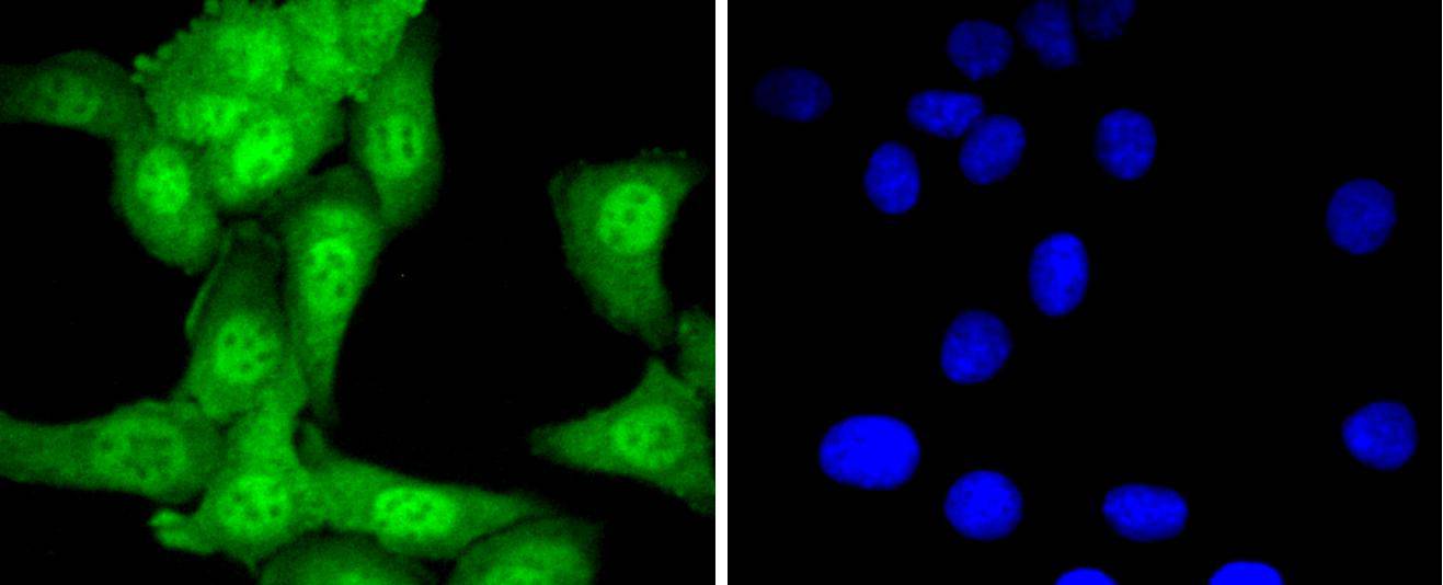
ICC staining UBA1 in Hela cells (green). The nuclear counter stain is DAPI (blue). Cells were fixed in paraformaldehyde, permeabilised with 0.25% Triton X100/PBS.
ICC staining UBA1 in SKOV-3 cells (green). The nuclear counter stain is DAPI (blue). Cells were fixed in paraformaldehyde, permeabilised with 0.25% Triton X100/PBS.
ICC staining UBA1 in A549 cells (green). The nuclear counter stain is DAPI (blue). Cells were fixed in paraformaldehyde, permeabilised with 0.25% Triton X100/PBS.
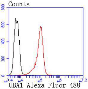
Flow cytometric analysis of K562 cells with UBA1 antibody at 1/50 dilution (red) compared with an unlabelled control (cells without incubation with primary antibody; black). Alexa Fluor 488-conjugated goat anti rabbit IgG was used as the secondary antibody.
The ubiquitin activating enzyme E1 (UBE1) catalyzes the first step in ubiquitin conjugation to mark cellular proteins for degradation. Specifically, UBE1 functions to adenylate the C-terminal glycine residue of ubiquitin, a reaction that is ATP-dependent and is proceeded by the formation of a thiolester bond with a cysteine residue of UBE1. The UBE1-activated ubiquitin is then transferred to a ubiquitin conjugated enzyme, which donates the ubiquitin residue to target substrates. The UBE1 gene is an example of an X-Y homologous gene, which is X-linked with a distinct Y-linked gene in many mammals. However, no UBE1 homolog is detectable on the human Y chromosome. UBE1 is thought to escape X inactivation in humans.

If you have published an article using product 49197, please notify us so that we can cite your literature.

NOTE

Application

  • WBWestern Blotting
  • IHCImmunohistochemistry
  • IFImmunofluorescence
  • ICCImmunocytochemistry
  • FCFlow Cytometry
  • IPImmunoprecipitation
  • EELISA
  • DBDot Blotting
  • ChIPChromatin Immunoprecipitation
  • GICAGold Immunochromatography Assay
  • NCNegative Control

Species Reactivity

  • HuHuman
  • MsMouse
  • RtRat
  • DmDrosophila melanogaster
  • CCaenorhabditis elegans
  • MkMonkey
  • RbRabbit
  • BBovine
  • DDog
  • PPig
  • HmHamster
  • ChHmChinese Hamster
  • ChkChicken
  • ShpSheep
;