Welcome to Sabbiotech!     
Email:info@signalwayantibody.com
Location: Home > Recombinant Rabbit Monoclonal Antibodies > Collagen VI Rabbit mAb

Collagen VI Rabbit mAb#49207

Availability: Yes
Total Amount:$
Product Detail

Product NameCollagen VI Rabbit mAb

Clone No.SD83-03

Host SpeciesRecombinant Rabbit

Clonality Monoclonal

PurificationProA affinity purified

ApplicationsWB, ICC/IF, IHC

Species ReactivityHu, Ms, Rt

Immunogen Descrecombinant protein

ConjugateUnconjugated

Other NamesAlpha 1 (VI) chain (61 AA) antibody
CO6A1_HUMAN antibody
COL6A1 antibody
COL6A2 antibody
COL6A3 antibody
Collagen alpha 2(VI) chain antibody
Collagen alpha 3(VI) chain antibody
Collagen alpha-1(VI) chain antibody
Collagen type VI alpha 1 antibody
Collagen type VI alpha 2 antibody
Collagen type VI alpha 3 antibody
Collagen VI alpha 1 polypeptide antibody
Collagen VI alpha 2 polypeptide antibody
Collagen VI alpha 3 polypeptide antibody
CollagenVI antibody
Human mRNA for collagen VI alpha 2 C terminal globular domain antibody
OPLL antibody
PP3610 antibody

Accession NoSwiss-Prot#:P12109

Uniprot P12109

Gene ID 1291;

Calculated MW134/109 kDa

Formulation1*TBS (pH7.4), 1%BSA, 40%Glycerol. Preservative: 0.05% Sodium Azide.

StorageStore at -20˚C

Application Details
WB: 1:1,000-1:2,000
IHC: 1:50-1:200
ICC: 1:100-1:500

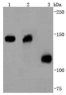
Western blot analysis of Collagen VI on different lysates using anti-Collagen VI antibody at 1/1,000 dilution. Positive control:
Lane 1: Mouse heart
Lane 2: Human heart
Lane 3: Human kidney
Immunohistochemical analysis of paraffin-embedded human lung tissue using anti-Collagen VI antibody. Counter stained with hematoxylin.
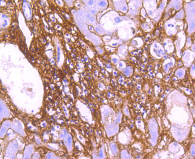
Immunohistochemical analysis of paraffin-embedded human liver tissue using anti-Collagen VI antibody. Counter stained with hematoxylin.
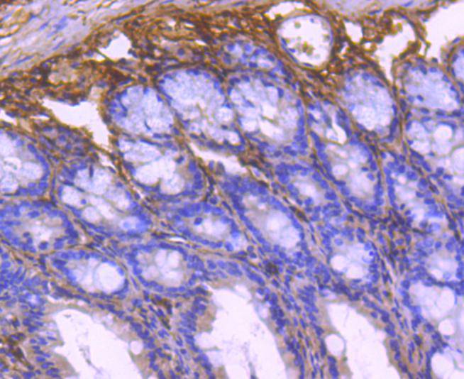
Immunohistochemical analysis of paraffin-embedded mouse colon tissue using anti-Collagen VI antibody. Counter stained with hematoxylin.
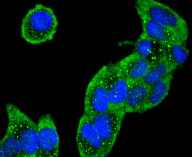
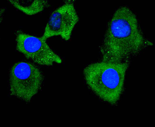
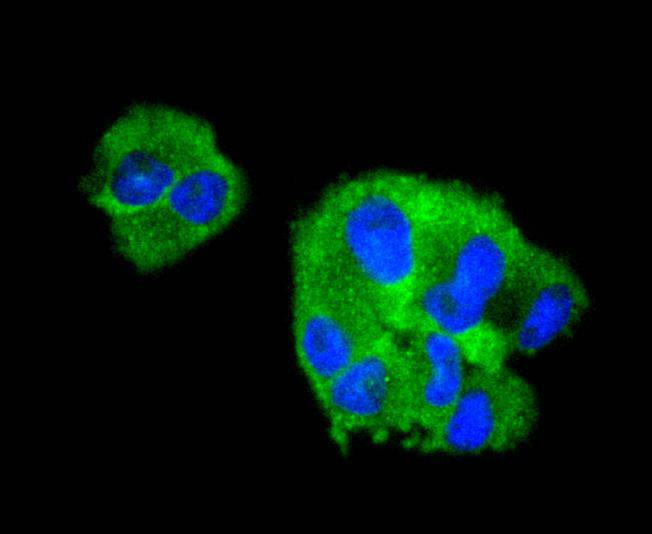
ICC staining Collagen VI in Hela cells (green). The nuclear counter stain is DAPI (blue). Cells were fixed in paraformaldehyde, permeabilised with 0.25% Triton X100/PBS.
ICC staining Collagen VI in A549 cells (green). The nuclear counter stain is DAPI (blue). Cells were fixed in paraformaldehyde, permeabilised with 0.25% Triton X100/PBS.
ICC staining Collagen VI in HepG2 cells (green). The nuclear counter stain is DAPI (blue). Cells were fixed in paraformaldehyde, permeabilised with 0.25% Triton X100/PBS.
ICC staining Collagen VI in RH-35 cells (green). The nuclear counter stain is DAPI (blue). Cells were fixed in paraformaldehyde, permeabilised with 0.25% Triton X100/PBS.
This antibody is well suited to detect extracellular matrix proteins in normal as well as disease state tissues. Disruption of tissue organization is the hallmark of neoplasia. Malignant lesions can be distinguished from benign by examining the breakdown of basement membranes and loss of 3-dimensional architecture. Malignant cells are presumed to use matrix metalloproteases to degrade barriers created by the extracellular matrix which then allows metastasis to occur. Collagenases, stomelysins and gelatinases can collectively degrade all of the various components of the extracellular matrix, including fibrillar and non-fibrillar collagens and basement membrane glycoproteins.

If you have published an article using product 49207, please notify us so that we can cite your literature.

NOTE

Application

  • WBWestern Blotting
  • IHCImmunohistochemistry
  • IFImmunofluorescence
  • ICCImmunocytochemistry
  • FCFlow Cytometry
  • IPImmunoprecipitation
  • EELISA
  • DBDot Blotting
  • ChIPChromatin Immunoprecipitation
  • GICAGold Immunochromatography Assay
  • NCNegative Control

Species Reactivity

  • HuHuman
  • MsMouse
  • RtRat
  • DmDrosophila melanogaster
  • CCaenorhabditis elegans
  • MkMonkey
  • RbRabbit
  • BBovine
  • DDog
  • PPig
  • HmHamster
  • ChHmChinese Hamster
  • ChkChicken
  • ShpSheep
;