Product Detail
Product Name14-3-3 Theta Rabbit mAb
Clone No.SD084-02
Host SpeciesRecombinant Rabbit
Clonality Monoclonal
PurificationProA affinity purified
ApplicationsWB, ICC/IF, FC
Species ReactivityHu, Ms, Rt
Immunogen Descrecombinant protein
ConjugateUnconjugated
Other Names14-3-3 antibody 14-3-3 protein T-cell antibody 14-3-3 protein tau antibody 14-3-3 protein theta antibody 1C5 antibody HS1 antibody theta polypeptide antibody tyrosine 3-monooxygenase/tryptophan 5-monooxygenase activation protein antibody tyrosine 3-monooxygenase/tryptophan 5-monooxygenase activation protein, theta isoform antibody tyrosine 3-monooxygenase/tryptophan 5-monooxygenase activation protein, theta polypeptide antibody
Accession NoSwiss-Prot#:P27348
Uniprot
P27348
Gene ID
10971;
Calculated MW28 kDa
Formulation1*TBS (pH7.4), 1%BSA, 40%Glycerol. Preservative: 0.05% Sodium Azide.
StorageStore at -20˚C
Application Details
WB: 1:1,000-5,000
ICC: 1:100-1:500
FC: 1:50-1:100
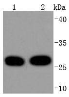
Western blot analysis of 14-3-3 Theta on different lysates using anti-14-3-3 Theta antibody at 1/1,000 dilution. Positive control: Lane 1: Hela Lane 2: MCF-7
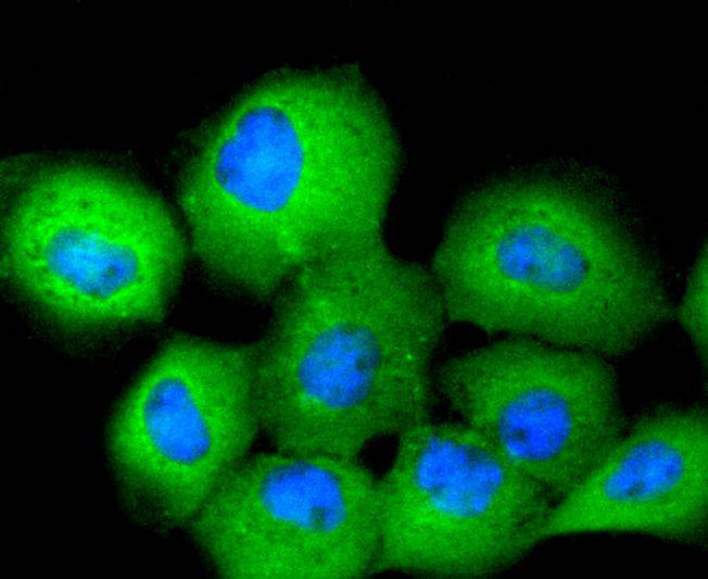
ICC staining 14-3-3 Theta in Hela cells (green). The nuclear counter stain is DAPI (blue). Cells were fixed in paraformaldehyde, permeabilised with 0.25% Triton X100/PBS.
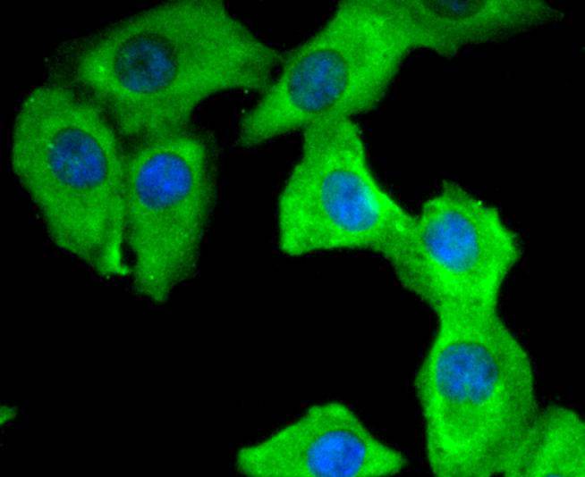
ICC staining 14-3-3 Theta in A431 cells (green). The nuclear counter stain is DAPI (blue). Cells were fixed in paraformaldehyde, permeabilised with 0.25% Triton X100/PBS.
ICC staining 14-3-3 Theta in A549 cells (green). The nuclear counter stain is DAPI (blue). Cells were fixed in paraformaldehyde, permeabilised with 0.25% Triton X100/PBS.
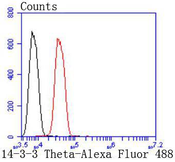
Flow cytometric analysis of SH-SY-5Y cells with 14-3-3 Theta antibody at 1/50 dilution (red) compared with an unlabelled control (cells without incubation with primary antibody; black). Alexa Fluor 488-conjugated goat anti rabbit IgG was used as the secondary antibody.
14-3-3 proteins regulate many cellular processes relevant to cancer biology, notably apoptosis, mitogenic signaling and cell-cycle checkpoints. Seven isoforms comprise this family of signaling intermediates, denoted 14-3-3 β, γ, ε, ζ, η, θ and . 14-3-3 proteins form dimers that present two binding sites for ligand proteins, thereby bringing together two proteins that may not otherwise associate. These ligands largely share a 14-3-3 consensus binding motif and exhibit serine/threonine phosphorylation. 14-3-3 proteins function in broad regulation of these ligand proteins, by cytoplasmic sequestration, occupation of interaction domains and import/export sequences, prevention of degradation, activation/repression of enzymatic activity and facilitation of protein modification, and thus loss of expression contributes to a vast array of pathogenic cellular activities.
If you have published an article using product 49213, please notify us so that we can cite your literature.


 Yes
Yes


