Welcome to Sabbiotech!     
Email:info@signalwayantibody.com
Location: Home > Recombinant Rabbit Monoclonal Antibodies > LC3B Rabbit mAb

LC3B Rabbit mAb#49277

Availability: Yes
Total Amount:$
Product Detail

Product NameLC3B Rabbit mAb

Clone No.JJ090-6

ClonalityMonoclonal

PurificationProA affinity purified

ApplicationsWB, IP

Species ReactivityHu, Ms, Rt

Immunogen Descrecombinant protein

ConjugateUnconjugated

Other NamesATG8F antibody
Autophagy-related protein LC3 B antibody
Autophagy-related ubiquitin-like modifier LC3 B antibody
LC3B antibody
LC3II antibody
MAP1 light chain 3 like protein 2 antibody
MAP1 light chain 3-like protein 2 antibody
MAP1A/1BLC3 antibody
MAP1A/MAP1B LC3 B antibody
MAP1A/MAP1B light chain 3 B antibody
MAP1ALC3 antibody
MAP1LC3B a antibody
Map1lc3b antibody
Microtubule associated protein 1 light chain 3 beta antibody
Microtubule-associated protein 1 light chain 3 beta antibody
Microtubule-associated proteins 1A/1B light chain 3B antibody
MLP3B_HUMAN antibody

Accession NoSwiss-Prot#:Q9GZQ8

Uniprot Q9GZQ8

Gene ID 81631;

Calculated MW14 kDa

Formulation1*TBS (pH7.4), 1%BSA, 40%Glycerol. Preservative: 0.05% Sodium Azide.

StorageStore at -20˚C

Application Details

WB: 1:1,000-1:2,000


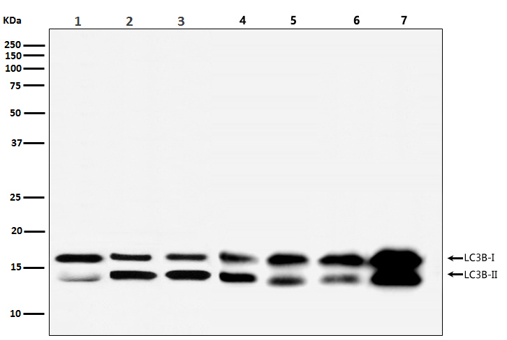
All lanes :LC3B Rabbit mAb at 1/2k dilution
Lane 1 : Human brain lysate
Lane 2 : RAW 264.7 cell lysate
Lane 3 : C6 cell lysate
Lane 4 : 293 cell lysate
Lane 5 : THP-1 cell lysate
Lane 6 : A431 cell lysate
Lane 7 : SH-SY5Y cell lysate
Secondary
All lanes : Goat Anti-Rabbit IgG H&L (HRP) at 1/10000 dilution
Predicted band size: 14,16 kDa
Observed band size: 14,16 kDa
Exposure time: 130 seconds

Microtubule-associated proteins (MAPs) regulate microtubule stability and play critical roles in neuronal development and in maintaining the balance between neuronal plasticity and rigidity. MAP-light chain 3 beta (MAP-LC3β) and MAP-light chain 3 alpha (MAP-LC3α) are subunits of both MAP1A and MAP1B. MAP-LC3β, a homolog of Apg8p, is essential for autophagy and associated to the autophagosome membranes after processing. Two forms of LC3β, the cytosolic LC3-I and the membrane-bound LC3-II, are produced post-translationally. LC3-I is formed by the removal of the C-terminal 22 amino acids from newly synthesized LC3β, followed by the conversion of a fraction of LC3-I into LC3-II. LC3 enhances fibronectin mRNA translation in ductus arteriosus cells through association with 60S ribosomes and binding to an AU-rich element in the 3 untranslated region of fibronectin mRNA. This facilitates sorting of fibronectin mRNA onto rough endoplasmic reticulum and translation. MAP LC3β may also be involved in formation of autophagosomal vacuoles. It is expressed primarily in heart, testis, brain and skeletal muscle.

If you have published an article using product 49277, please notify us so that we can cite your literature.

NOTE

Application

  • WBWestern Blotting
  • IHCImmunohistochemistry
  • IFImmunofluorescence
  • ICCImmunocytochemistry
  • FCFlow Cytometry
  • IPImmunoprecipitation
  • EELISA
  • DBDot Blotting
  • ChIPChromatin Immunoprecipitation
  • GICAGold Immunochromatography Assay
  • NCNegative Control

Species Reactivity

  • HuHuman
  • MsMouse
  • RtRat
  • DmDrosophila melanogaster
  • CCaenorhabditis elegans
  • MkMonkey
  • RbRabbit
  • BBovine
  • DDog
  • PPig
  • HmHamster
  • ChHmChinese Hamster
  • ChkChicken
  • ShpSheep
;