Product Detail
Product NameCystatin C Rabbit mAb
Clone No.JJ09-16
Host SpeciesRecombinant Rabbit
Clonality Monoclonal
PurificationProA affinity purified
ApplicationsWB, IHC
Species ReactivityHu, Ms, Rt
Immunogen Descrecombinant protein
ConjugateUnconjugated
Other NamesAD 8 antibody AD8 antibody Amyloid angiopathy and cerebral hemorrhage antibody ARMD11 antibody bA218C14.4 (cystatin C) antibody bA218C14.4 antibody Cst 3 antibody Cst3 antibody CST3 protein antibody Cystatin 3 antibody Cystatin-3 antibody Cystatin-C antibody Cystatin3 antibody CystatinC antibody CYTC_HUMAN antibody Epididymis secretory protein Li 2 antibody Gamma trace antibody Gamma-trace antibody HCCAA antibody HEL S 2 antibody MGC117328 antibody Neuroendocrine basic polypeptide antibody Post gamma globulin antibody Post-gamma-globulin antibody
Accession NoSwiss-Prot#:P01034
Uniprot
P01034
Gene ID
1471;
Calculated MW16 kDa
Formulation1*TBS (pH7.4), 1%BSA, 40%Glycerol. Preservative: 0.05% Sodium Azide.
StorageStore at -20˚C
Application Details
WB: 1:1,000-1:2,000
IHC: 1:50-1:200
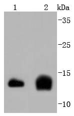
Western blot analysis of Cystatin C on different lysates using anti-Cystatin C antibody at 1/1,000 dilution. Positive control: Lane 1: Hela Lane 2: Mouse spleen
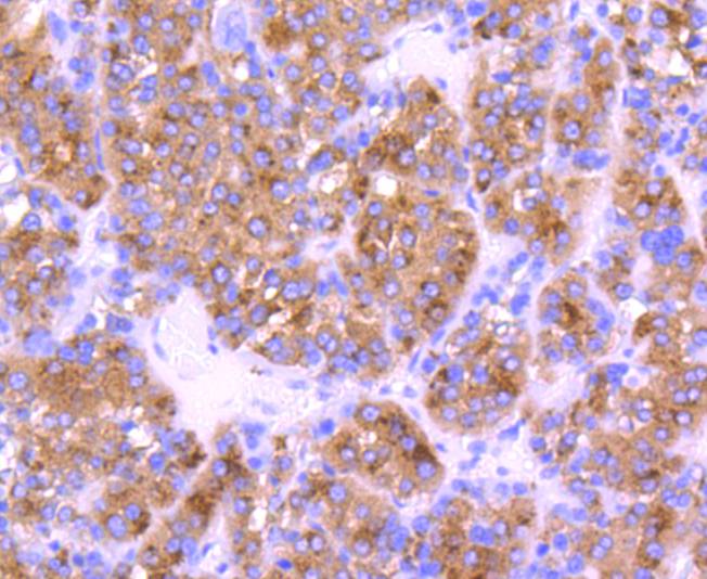
Immunohistochemical analysis of paraffin-embedded human liver cancer tissue using anti-Cystatin C antibody. Counter stained with hematoxylin.
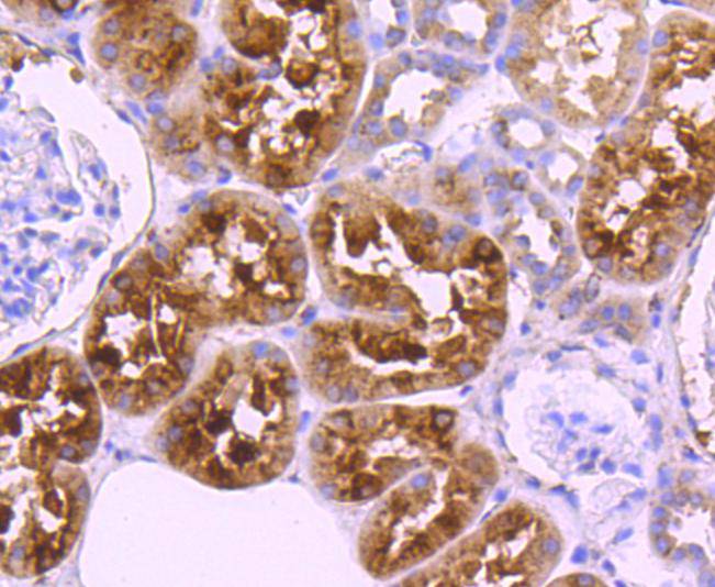
Immunohistochemical analysis of paraffin-embedded human kidney tissue using anti-Cystatin C antibody. Counter stained with hematoxylin.
Immunohistochemical analysis of paraffin-embedded mouse brain tissue using anti-Cystatin C antibody. Counter stained with hematoxylin.
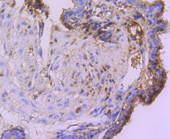
Immunohistochemical analysis of paraffin-embedded mouse placenta tissue using anti-Cystatin C antibody. Counter stained with hematoxylin.
Immunohistochemical analysis of paraffin-embedded mouse kidney tissue using anti-Cystatin C antibody. Counter stained with hematoxylin.
Cystatin C is a cysteine (thiol) protease inhibitor that belongs to the type II cystatin gene superfamily and is the most abundant extracellular inhibitor of cysteine proteases. Cystatin C is a constitutively secreted, amyloidogenic protein, which forms a two-fold symmetric dimer and modulates both cysteine protease activity and the expression of class II MHC molecules. Expression of cystatin C is an indicator of kidney function and glomerular filtration rate. Mutations in the cystatin C gene can lead to protein aggregates, which are implicated in hereditary amyloid angiopathy (HCCAA) and cerebral hemorrhage. Although both wild-type and mutant cystatin C are capable of forming concentration dependent inactive dimers, mutant cystatin C dimerizes at lower concentrations and is more susceptible to serine proteases, which may facilitate aggregation. In neuronal cells, oxidative stress stimulates expression of cystatin C, which may positively regulate apoptosis.
If you have published an article using product 49285, please notify us so that we can cite your literature.


 Yes
Yes


