Welcome to Sabbiotech!     
Email:info@signalwayantibody.com
Location: Home > Recombinant Rabbit Monoclonal Antibodies > DUSP1 Rabbit mAb

DUSP1 Rabbit mAb#49296

Availability: Yes
Total Amount:$
Product Detail

Product NameDUSP1 Rabbit mAb

Clone No.JJ0930

Host SpeciesRecombinant Rabbit

ClonalityMonoclonal

PurificationProA affinity purified

ApplicationsWB, IHC, IP, FC

Species ReactivityHu, Ms, Rt

Immunogen Descrecombinant protein

ConjugateUnconjugated

Other NamesCL 100 antibody
CL100 antibody
Dual Specificity Phosphatase 1 antibody
Dual specificity protein phosphatase 1 antibody
Dual specificity protein phosphatase hVH1 antibody
DUS1_HUMAN antibody
DUSP 1 antibody
Dusp1 antibody
HVH1 antibody
MAP kinase phosphatase 1 antibody
Mitogen-activated protein kinase phosphatase 1 antibody
MKP-1 antibody
MKP1 antibody
Protein tyrosine phosphatase CL100 antibody
Protein-tyrosine phosphatase CL100 antibody
PTPN10 antibody
Serine/threonine specific protein phosphatase antibody
VH1 antibody

Accession NoSwiss-Prot#:P28562

Uniprot P28562

Gene ID 1843;

Calculated MW40 kDa

Formulation1*TBS (pH7.4), 1%BSA, 40%Glycerol. Preservative: 0.05% Sodium Azide.

StorageStore at -20˚C

Application Details
WB: 1:1,000
IHC: 1:50-1:200


FC: 1:50-1:100
Western blot analysis of DUSP1 on mouse spleen lysates using anti-DUSP1 antibody at 1/1,000 dilution.
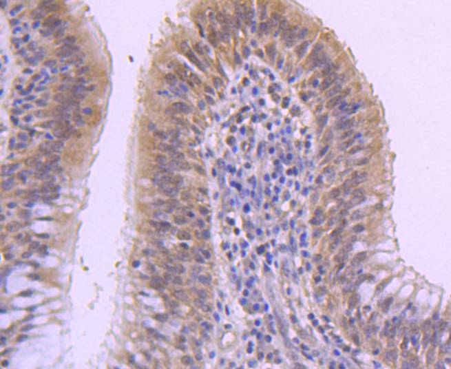
Immunohistochemical analysis of paraffin-embedded human lung cancer tissue using anti-DUSP1 antibody. Counter stained with hematoxylin.
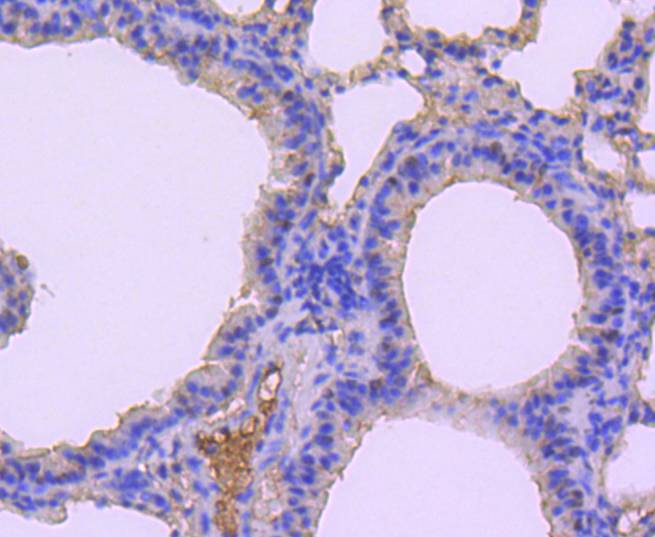
Immunohistochemical analysis of paraffin-embedded mouse lung tissue using anti-DUSP1 antibody. Counter stained with hematoxylin.
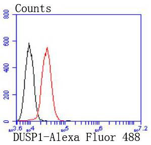
Flow cytometric analysis of HepG2 cells with DUSP1 antibody at 1/50 dilution (red) compared with an unlabelled control (cells without incubation with primary antibody; black). Alexa Fluor 488-conjugated goat anti rabbit IgG was used as the secondary antibody
A key element in the pathway involved in the transduction of signals from activated protein-tyrosine kinase transmembrane receptors has been identified as the family of mitogen-activated protein kinases (MAP kinases). The most well known of these Ser/Thr kinases are ERK 1 and ERK 2. Mitogenic stimulation of cells triggers the activation of MAP kinases through phosphorylation of both tyrosyl (Y185) and threonyl (T183) residues. Phosphorylation of the T183 and Y185 ERK regulatory site is mediated by MAP kinase (MEK), which in turn is regulated by the proto-oncogene product Raf. Two highly related phosphatases, designated MKP-1 and MKP-2, exhibit 59% sequence identity at the amino acid level and oppose the action of MEK by downregulating the kinase activity of ERK 1 and ERK 2. MAP kinase phosphatase-1 and -2 proteins function by dephosphorylating ERK 1 and ERK 2 at their T-E-Y regulatory motif. An additional phosphatase encoded by the DUSP2 gene, designated PAC-1, also functions to downregulate ERK 1 and ERK 2 kinase activity. PAC-1 is a nuclear protein whose expression is strongly induced in response to mitogen.

If you have published an article using product 49296, please notify us so that we can cite your literature.

NOTE

Application

  • WBWestern Blotting
  • IHCImmunohistochemistry
  • IFImmunofluorescence
  • ICCImmunocytochemistry
  • FCFlow Cytometry
  • IPImmunoprecipitation
  • EELISA
  • DBDot Blotting
  • ChIPChromatin Immunoprecipitation
  • GICAGold Immunochromatography Assay
  • NCNegative Control

Species Reactivity

  • HuHuman
  • MsMouse
  • RtRat
  • DmDrosophila melanogaster
  • CCaenorhabditis elegans
  • MkMonkey
  • RbRabbit
  • BBovine
  • DDog
  • PPig
  • HmHamster
  • ChHmChinese Hamster
  • ChkChicken
  • ShpSheep
;