Welcome to Sabbiotech!     
Email:info@signalwayantibody.com
Location: Home > Recombinant Rabbit Monoclonal Antibodies > 67kDa Laminin Receptor Rabbit mAb

67kDa Laminin Receptor Rabbit mAb#49319

Availability: Yes
Total Amount:$
Product Detail

Product Name67kDa Laminin Receptor Rabbit mAb

Clone No.JF0955

ClonalityMonoclonal

PurificationProA affinity purified

ApplicationsWB, ICC/IF, IHC, IP, FC

Species ReactivityHu, Ms, Rt

Immunogen Descrecombinant protein

ConjugateUnconjugated

Other Names34/67 kDa laminin receptor antibody
37 kDa laminin receptor precursor antibody
37/67 kDa laminin receptor antibody
37LRP antibody
40S ribosomal protein SA antibody
67 kDa laminin receptor antibody
67LR antibody
Colon carcinoma laminin binding protein antibody
Colon carcinoma laminin-binding protein antibody
LAMBR antibody
Laminin receptor 1 antibody
Laminin-binding protein precursor p40 antibody
LAMR 1 antibody
LamR antibody
LAMR1 antibody
LBP antibody
LBP/p40 antibody
LRP antibody
LRP/LR antibody
Multidrug resistance associated protein MGr1 Ag antibody
Multidrug resistance associated protein MGr1Ag antibody
Multidrug resistance-associated protein MGr1-Ag antibody
NEM/1CHD4 antibody
p40 antibody
Ribosomal Protein SA antibody
rpsA antibody
RSSA_HUMAN antibody
SA antibody

Accession NoSwiss-Prot#:P08865

Uniprot P08865

Gene ID 3921;

Calculated MW40 kDa

Concentration1 mg/ml

Formulation1*TBS (pH7.4), 1%BSA, 40%Glycerol. Preservative: 0.05% Sodium Azide.

StorageStore at -20˚C

Application Details

WB: 1:1,000-1:2,000
IHC: 1:50-1:200
ICC: 1:100-1:500

FC: 1:50-1:100

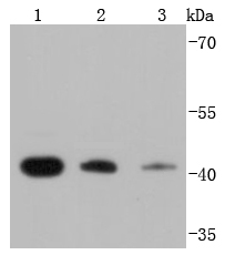
Western blot analysis of RPSA on different lysates using anti-RPSA antibody at 1/1,000 dilution. Positive control:
Lane 1: K562
Lane 2: HepG2
Lane 3: A431
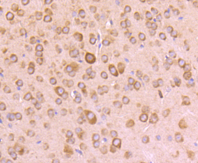
Immunohistochemical analysis of paraffin-embedded mouse brain tissue using anti-RPSA antibody. Counter stained with hematoxylin.
Immunohistochemical analysis of paraffin-embedded mouse kidney tissue using anti-RPSA antibody. Counter stained with hematoxylin.
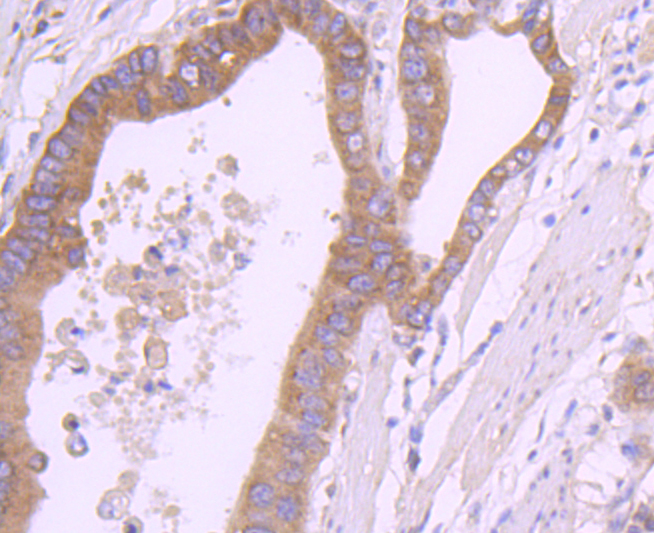
Immunohistochemical analysis of paraffin-embedded human gastric carcinoma tissue using anti-RPSA antibody. Counter stained with hematoxylin.
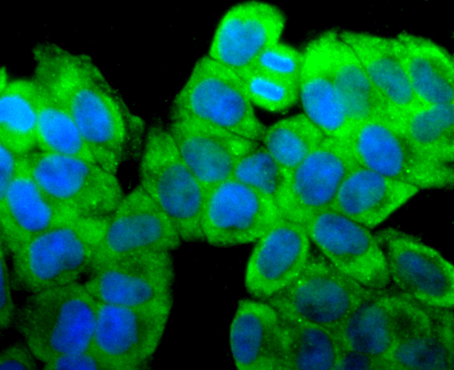
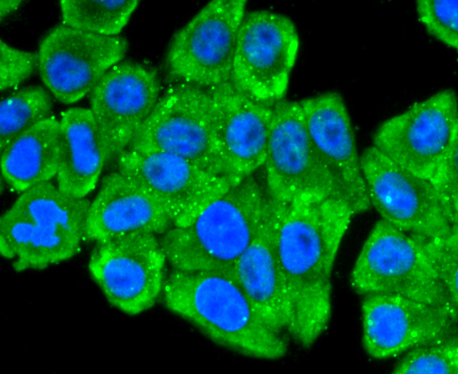
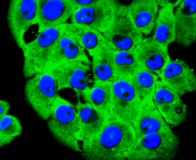
ICC staining RPSA in Hela cells (green). The nuclear counter stain is DAPI (blue). Cells were fixed in paraformaldehyde, permeabilised with 0.25% Triton X100/PBS.
ICC staining RPSA in MCF-7 cells (green). The nuclear counter stain is DAPI (blue). Cells were fixed in paraformaldehyde, permeabilised with 0.25% Triton X100/PBS.
ICC staining RPSA in HepG2 cells (green). The nuclear counter stain is DAPI (blue). Cells were fixed in paraformaldehyde, permeabilised with 0.25% Triton X100/PBS.
ICC staining RPSA in RH-35 cells (green). The nuclear counter stain is DAPI (blue). Cells were fixed in paraformaldehyde, permeabilised with 0.25% Triton X100/PBS.
Flow cytometric analysis of MCF-7 cells with RPSA antibody at 1/50 dilution (red) compared with an unlabelled control (cells without incubation with primary antibody; black). Alexa Fluor 488-conjugated goat anti rabbit IgG was used as the secondary antibody.

Laminin receptor (Laminin-R) has a heterodimeric structure similar to that of receptors for other extracellular matrix proteins such as Fibronectin and Vitronectin. Incorporation of Laminin-R into lysosomal membranes makes it possible for lysosomes to attach to surfaces coated with Laminin. This and other properties identify Laminin-R as a member of the integrin family of cell adhesion receptors. The Laminin-R precursor is a polypeptide whose expression is consistently upregulated in aggressive carcinoma. The precursor, which is also identified as p40 ribosome-associated protein, appears to be a multifunctional protein involved in the translational machinery. Laminin-R (also known as colon carcinoma laminin-binding protein) and is found at nine-fold higher levels in colon carcinoma than in adjacent normal colonic epithelium. Additionally, the level of the Laminin-R is higher in the lung cancer cell line than in the lung cell line.

If you have published an article using product 49319, please notify us so that we can cite your literature.

NOTE

Application

  • WBWestern Blotting
  • IHCImmunohistochemistry
  • IFImmunofluorescence
  • ICCImmunocytochemistry
  • FCFlow Cytometry
  • IPImmunoprecipitation
  • EELISA
  • DBDot Blotting
  • ChIPChromatin Immunoprecipitation
  • GICAGold Immunochromatography Assay
  • NCNegative Control

Species Reactivity

  • HuHuman
  • MsMouse
  • RtRat
  • DmDrosophila melanogaster
  • CCaenorhabditis elegans
  • MkMonkey
  • RbRabbit
  • BBovine
  • DDog
  • PPig
  • HmHamster
  • ChHmChinese Hamster
  • ChkChicken
  • ShpSheep
;