Product Detail
Product NameLYVE1 Rabbit mAb
Clone No.JF0979
Host SpeciesRecombinant Rabbit
Clonality Monoclonal
PurificationProA affinity purified
ApplicationsWB, ICC/IF, IHC, FC
Species ReactivityHu, Ms, Rt
Immunogen Descrecombinant protein
ConjugateUnconjugated
Other NamesCell surface retention sequence-binding protein 1 antibody CRSBP 1 antibody CRSBP-1 antibody CRSBP1 antibody extracellular link domain containing 1 antibody extracellular link domain-containing 1 antibody Extracellular link domain-containing protein 1 antibody HAR antibody Hyaluronic acid receptor antibody Lymphatic endothelium specific hyaluronan receptor antibody lymphatic vessel endothelial hyaluronan receptor 1 antibody Lymphatic vessel endothelial hyaluronic acid receptor 1 antibody LYVE 1 antibody LYVE-1 antibody LYVE1 antibody LYVE1_HUMAN antibody XLKD1 antibody
Accession NoSwiss-Prot#:Q9Y5Y7
Uniprot
Q9Y5Y7
Gene ID
10894;
Calculated MW35 kDa
Formulation1*TBS (pH7.4), 1%BSA, 40%Glycerol. Preservative: 0.05% Sodium Azide.
StorageStore at -20˚C
Application Details
WB: 1:1,000
IHC: 1:50-1:200
ICC: 1:50-1:200
FC: 1:50-1:100
Western blot analysis of LYVE1 on MCF-7 lysates using anti-LYVE1 antibody at 1/1,000 dilution.
Immunohistochemical analysis of paraffin-embedded human spleen tissue using anti-LYVE1 antibody. Counter stained with hematoxylin.
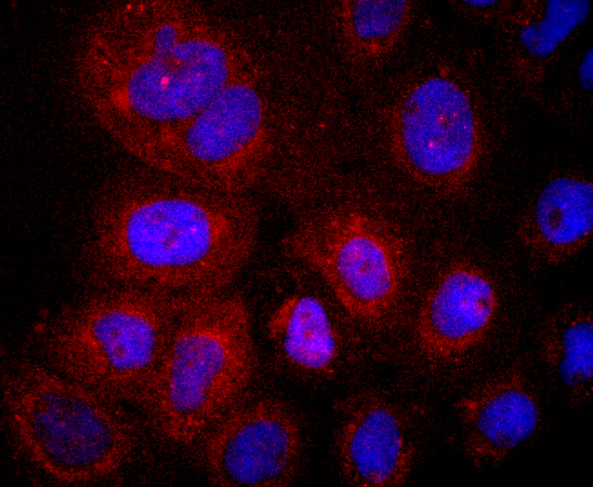
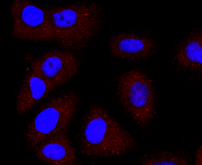
ICC staining LYVE1 in A431 cells (red). The nuclear counter stain is DAPI (blue). Cells were fixed in paraformaldehyde, permeabilised with 0.25% Triton X100/PBS.
ICC staining LYVE1 in HUVEC cells (red). The nuclear counter stain is DAPI (blue). Cells were fixed in paraformaldehyde, permeabilised with 0.25% Triton X100/PBS.
ICC staining LYVE1 in SW480 cells (red). The nuclear counter stain is DAPI (blue). Cells were fixed in paraformaldehyde, permeabilised with 0.25% Triton X100/PBS.
Flow cytometric analysis of HUVEC cells with LYVE1 antibody at 1/50 dilution (red) compared with an unlabelled control (cells without incubation with primary antibody; black). Alexa Fluor 488-conjugated goat anti rabbit IgG was used as the secondary antibody
Lymphatic vessel endothelial hyaluronan receptor-1 (LYVE-1) is expressed on the cell surface as a protein that is reduced by glycosidase treatment. LYVE-1 is abundant in spleen, lymph node, heart, lung and fetal liver, and is less abundant in appendix, bone marrow, placenta, muscle and adult liver. Expression of LYVE-1 is largely restricted to endothelial cells lining lymphatic vessels and splenic sinusoidal endothelial cells. LYVE-1 binds to both soluble and immobilized hyaluronan (HA) with greater specificity than HCAM. Like HCAM, the LYVE-1 molecule binds both soluble and immobilized HA. However, unlike HCAM, the LYVE-1 molecule co-localizes with HA on the luminal face of the lymph vessel wall and is completely absent from blood vessels. Hence, LYVE-1 is the first lymph-specific HA receptor to be characterized and is a uniquely powerful marker for lymph vessels themselves. LYVE-1 is used as a marker to study tumor lymphangiogenesis, which is an important area of investigation.
If you have published an article using product 49343, please notify us so that we can cite your literature.


 Yes
Yes


