Product Detail
Product NameThymidine Kinase 1 Rabbit mAb
Clone No.JF0970
Host SpeciesRecombinant Rabbit
Clonality Monoclonal
PurificationProA affinity purified
ApplicationsWB, ICC/IF, IHC, IP, FC
Species ReactivityHu
Immunogen Descrecombinant protein
ConjugateUnconjugated
Other Namescytosolic antibody KITH_HUMAN antibody Thymidine kinase 1 antibody Thymidine kinase 1 soluble antibody Thymidine kinase 1 soluble isoform antibody Thymidine kinase antibody Thymidine kinase cytosolic antibody TK 1 antibody TK 2 antibody TK1 antibody Tk1a antibody Tk1b antibody TK2 antibody
Accession NoSwiss-Prot#:P04183
Uniprot
P04183
Gene ID
7083;
Calculated MW25 kDa
Formulation1*TBS (pH7.4), 1%BSA, 40%Glycerol. Preservative: 0.05% Sodium Azide.
StorageStore at -20˚C
Application Details
WB: 1:1,000
IHC: 1:50-1:200
ICC: 1:50-1:200
FC: 1:50-1:100
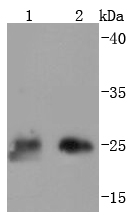
Western blot analysis of Thymidine Kinase 1 on different lysates using anti-Thymidine Kinase 1 antibody at 1/1,000 dilution. Positive control: Lane 1: 293T Lane 2: Hela
Immunohistochemical analysis of paraffin-embedded human tonsil tissue using anti-Thymidine Kinase 1 antibody. Counter stained with hematoxylin.
ICC staining Thymidine Kinase 1 in Hela cells (red). The nuclear counter stain is DAPI (blue). Cells were fixed in paraformaldehyde, permeabilised with 0.25% Triton X100/PBS.
ICC staining Thymidine Kinase 1 in NIH/3T3 cells (red). The nuclear counter stain is DAPI (blue). Cells were fixed in paraformaldehyde, permeabilised with 0.25% Triton X100/PBS.
ICC staining Thymidine Kinase 1 in SW480 cells (red). The nuclear counter stain is DAPI (blue). Cells were fixed in paraformaldehyde, permeabilised with 0.25% Triton X100/PBS.
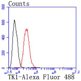
Flow cytometric analysis of Hela cells with Thymidine Kinase 1 antibody at 1/50 dilution (red) compared with an unlabelled control (cells without incubation with primary antibody; black). Alexa Fluor 488-conjugated goat anti rabbit IgG was used as the secondary antibody
Thymidine Kinase (TK1) is a highly conserved phosphotransferase that is present in most living cells. Thymidine Kinase catalyzes the phosphorylation reaction: deoxythymidine + ATP = deoxythymidine 5'-phosphate + ADP; it is thus involved in the reaction chain to introduce deoxythymidine into the DNA. Thymidine kinase is required for the action of many antiviral drugs, such as azidothymidine (AZT), and is is also used to select hybridoma cell lines in the production of monoclonal antibodies. Thymidine Kinase has many clinical applications as it is only present in anticipation of cell division. Because of this, Thymidine Kinase can be used as a proliferation marker in the diagnosis, treatment, and follow-up of malignant diseases, especially hematological malignancies. Thymidine Kinase may be observed as a monomer, dimer, trimer or tetramer.
If you have published an article using product 49345, please notify us so that we can cite your literature.


 Yes
Yes


