Welcome to Sabbiotech!     
Email:info@signalwayantibody.com
Location: Home > Recombinant Rabbit Monoclonal Antibodies > PDHA1 Rabbit mAb

PDHA1 Rabbit mAb#49389

Availability: Yes
Total Amount:$
Product Detail

Product NamePDHA1 Rabbit mAb

Clone No.JF996-0

Host SpeciesRecombinant Rabbit

Clonality Monoclonal

PurificationProA affinity purified

ApplicationsWB, ICC/IF, IHC, IP, FC

Species ReactivityHu, Ms, Rt

Immunogen Descrecombinant protein

ConjugateUnconjugated

Other NamesODPA_HUMAN antibody
PDH antibody
PDHA antibody
PDHA1 antibody
PDHCE1A antibody
PDHE1 A type I antibody
PDHE1-A type I antibody
PHE1A antibody
Pyruvate Dehydrogenase (lipoamide) alpha 1 antibody
Pyruvate dehydrogenase complex, E1 alpha polypeptide 1 antibody
Pyruvate Dehydrogenase E1 alpha antibody
Pyruvate dehydrogenase E1 component subunit alpha, somatic form, mitochondrial antibody

Accession NoSwiss-Prot#:P08559

Uniprot P08559

Gene ID 5160;

Calculated MW43 kDa

Formulation1*TBS (pH7.4), 1%BSA, 40%Glycerol. Preservative: 0.05% Sodium Azide.

StorageStore at -20˚C

Application Details
WB: 1:1,000-5,000
IHC: 1:50-1:200
ICC: 1:50-1:200

FC: 1:50-1:100
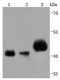
Western blot analysis of PDHA1 on different lysates using anti-PDHA1 antibody at 1/1,000 dilution. Positive control:
Lane 1: 293T
Lane 2: A431
Lane 3: Mouse heart
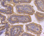
Immunohistochemical analysis of paraffin-embedded mouse stomach tissue using anti-PDHA1 antibody. Counter stained with hematoxylin.
Immunohistochemical analysis of paraffin-embedded human breast carcinoma tissue using anti-PDHA1 antibody. Counter stained with hematoxylin.
Immunohistochemical analysis of paraffin-embedded human kidney tissue using anti-PDHA1 antibody. Counter stained with hematoxylin.
Immunohistochemical analysis of paraffin-embedded mouse skeletal muscle tissue using anti-PDHA1 antibody. Counter stained with hematoxylin.
Immunohistochemical analysis of paraffin-embedded human lung cancer tissue using anti-PDHA1 antibody. Counter stained with hematoxylin.
Immunohistochemical analysis of paraffin-embedded mouse colon tissue using anti-PDHA1 antibody. Counter stained with hematoxylin.
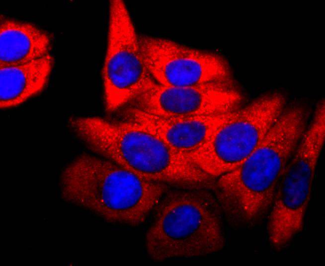
ICC staining PDHA1 in Hela cells (red). The nuclear counter stain is DAPI (blue). Cells were fixed in paraformaldehyde, permeabilised with 0.25% Triton X100/PBS.
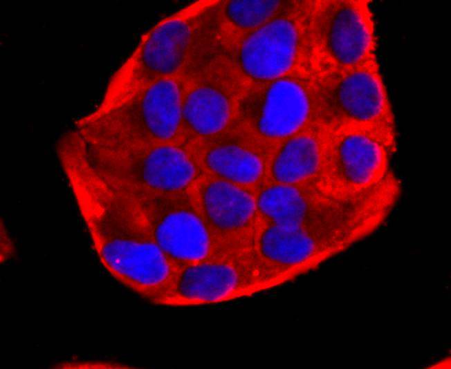
ICC staining PDHA1 in HepG2 cells (red). The nuclear counter stain is DAPI (blue). Cells were fixed in paraformaldehyde, permeabilised with 0.25% Triton X100/PBS.
ICC staining PDHA1 in SH-SY-5Y cells (red). The nuclear counter stain is DAPI (blue). Cells were fixed in paraformaldehyde, permeabilised with 0.25% Triton X100/PBS.
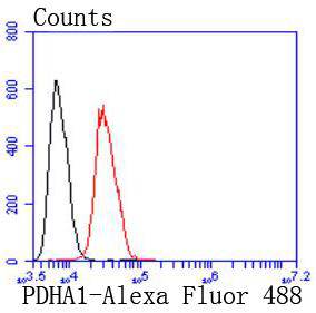
Flow cytometric analysis of Hela cells with PDHA1 antibody at 1/50 dilution (red) compared with an unlabelled control (cells without incubation with primary antibody; black). Alexa Fluor 488-conjugated goat anti rabbit IgG was used as the secondary antibody
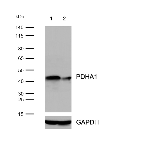
All lanes : PDHA1 epsilon Rabbit mAb at 1/1k dilution
Lane 1 : Wild-type HAP1 cell lysate
Lane 2 : PDHA1 knockout HAP1 cell lysate
Lysates/proteins at 20 µg per lane.
The pyruvate dehydrogenase (PDH) complex is a nuclear-encoded mitochondrial matrix enzyme complex that functions as the primary link between glycolysis and the tricarboxylic acid (TCA) cycle by catalyzing the irreversible conversion of pyruvate into acetyl-CoA. The E1 enzyme of the PDH complex is made up of a heterotetramer of two α and two β subunits. The E1-α subunit (PDH-E1α) contains the E1 active site and plays a key role in the function of the PDH complex. The PDH complex is regulated by phosphorylation and dephosphorylation of PDH-E1α. The gene encoding for PDH-E1α maps to chromosome Xp22.12, and a 20bp deletion in the last exon of this gene is sufficient to cause PDH deficiency, which causes a broad range of symptoms including the development of seizures, mental retardation and spasticity, as well as intermittent episodes of lactic acidosis associated with cerebellar ataxia.

If you have published an article using product 49389, please notify us so that we can cite your literature.

NOTE

Application

  • WBWestern Blotting
  • IHCImmunohistochemistry
  • IFImmunofluorescence
  • ICCImmunocytochemistry
  • FCFlow Cytometry
  • IPImmunoprecipitation
  • EELISA
  • DBDot Blotting
  • ChIPChromatin Immunoprecipitation
  • GICAGold Immunochromatography Assay
  • NCNegative Control

Species Reactivity

  • HuHuman
  • MsMouse
  • RtRat
  • DmDrosophila melanogaster
  • CCaenorhabditis elegans
  • MkMonkey
  • RbRabbit
  • BBovine
  • DDog
  • PPig
  • HmHamster
  • ChHmChinese Hamster
  • ChkChicken
  • ShpSheep
;