Product Detail
Product NameMiTF Rabbit mAb
Clone No.JF100-01
Host SpeciesRecombinant Rabbit
Clonality Monoclonal
PurificationProA affinity purified
ApplicationsWB, ICC, FC
Species ReactivityHu, Ms, Rt
Immunogen Descrecombinant protein
ConjugateUnconjugated
Other NamesBHLHE32 antibody bHLHe32 antibody Class E basic helix-loop-helix protein 32 antibody CMM8 antibody Homolog of mouse microphthalmia antibody Mi antibody Microphthalmia associated transcription factor antibody Microphthalmia, mouse, homolog of antibody Microphthalmia-associated transcription factor antibody MITF antibody MITF_HUMAN antibody mitfa antibody nacre antibody WS2 antibody WS2A antibody z3A.1 antibody
Accession NoSwiss-Prot#:O75030
Uniprot
O75030
Gene ID
4286;
Calculated MW55 kDa
Formulation1*TBS (pH7.4), 1%BSA, 40%Glycerol. Preservative: 0.05% Sodium Azide.
StorageStore at -20˚C
Application Details
WB: 1:1,000-1:2,000
ICC: 1:100-1:500
FC: 1:50-1:100
Western blot analysis of MiTF on PC-12 cells lysates using anti-MiTF antibody at 1/1,000 dilution.
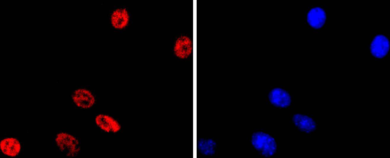
ICC staining MiTF in Hela cells (red). The nuclear counter stain is DAPI (blue). Cells were fixed in paraformaldehyde, permeabilised with 0.25% Triton X100/PBS.
ICC staining MiTF in A431 cells (red). The nuclear counter stain is DAPI (blue). Cells were fixed in paraformaldehyde, permeabilised with 0.25% Triton X100/PBS.
ICC staining MiTF in NIH/3T3 cells (red). The nuclear counter stain is DAPI (blue). Cells were fixed in paraformaldehyde, permeabilised with 0.25% Triton X100/PBS.
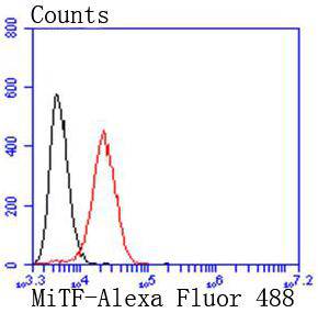
Flow cytometric analysis of SW480 cells with MiTF antibody at 1/50 dilution (red) compared with an unlabelled control (cells without incubation with primary antibody; black). Alexa Fluor 488-conjugated goat anti rabbit IgG was used as the secondary antibody.
MITF (microphthalmia-associated transcription factor) is a melanocytic nuclear protein that contains basic helix-loop-helix (HLH) and leucine zipper (LZ) domains. These protein motifs are frequently observed in other transcription factors and are particularly common to members of the Myc family. MITF can directly associate with DNA as a homodimer and is required for the development and differentiation of melanocytes. Its expression is upregulated by cAMP and cAMP-dependent pathways. MITF activates several different gene promoters by binding to their E-boxes. Tyrosinase, TRP1 and TRP2 are pigment synthesis genes activated by MITF. When MITF is phosphorylated on Ser73 (via the MAPK pathway), it associates with co-activators of the p300/CBP family and enhances transcription. MITF has several isoforms including MITF-M which is specifically expressed in melanocytes. In MITF-deficient mice there is a complete absence of melanocytes.
If you have published an article using product 49400, please notify us so that we can cite your literature.


 Yes
Yes


