Product Detail
Product Namealpha 1 Antitrypsin Rabbit mAb
Clone No.JF10-03
Host SpeciesRecombinant Rabbit
Clonality Monoclonal
PurificationProA affinity purified
ApplicationsWB, ICC/IF, IHC, IP
Species ReactivityHu
Immunogen Descrecombinant protein
ConjugateUnconjugated
Other NamesA1A antibody A1AT antibody A1AT_HUMAN antibody AAT antibody Alpha 1 antiproteinase antibody Alpha 1 antitrypsin antibody Alpha 1 antitrypsin null antibody Alpha 1 protease inhibitor antibody Alpha-1 protease inhibitor antibody Alpha-1-antiproteinase antibody alpha1 proteinase inhibitor antibody Alpha1AT antibody Dom1 antibody PI antibody PI1 antibody PRO2275 antibody Serine (or cysteine) proteinase inhibitor clade A member 1 antibody Serine protease inhibitor 1-1 antibody Serine protease inhibitor A1a antibody Serpin A1 antibody Serpin A1a antibody Serpin peptidase inhibitor clade A member 1 antibody Serpina1 antibody Short peptide from AAT antibody SPAAT antibody Spi1-1 antibody
Accession NoSwiss-Prot#:P01009
Uniprot
P01009
Gene ID
5265;
Calculated MW47 kDa
Formulation1*TBS (pH7.4), 1%BSA, 40%Glycerol. Preservative: 0.05% Sodium Azide.
StorageStore at -20˚C
Application Details
WB: 1:1,000-5,000
IHC: 1:50-1:200
ICC: 1:50-1:200
Western blot analysis of alpha 1 Antitrypsin on human spleen lysates using anti-alpha 1 Antitrypsin antibody at 1/1,000 dilution.
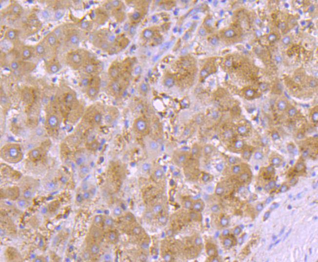
Immunohistochemical analysis of paraffin-embedded human liver tissue using anti-alpha 1 Antitrypsin antibody. Counter stained with hematoxylin.
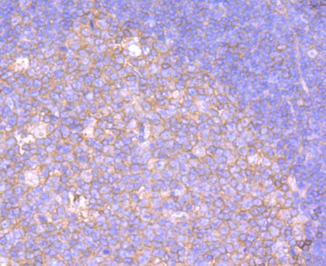
Immunohistochemical analysis of paraffin-embedded human spleen tissue using anti-alpha 1 Antitrypsin antibody. Counter stained with hematoxylin.
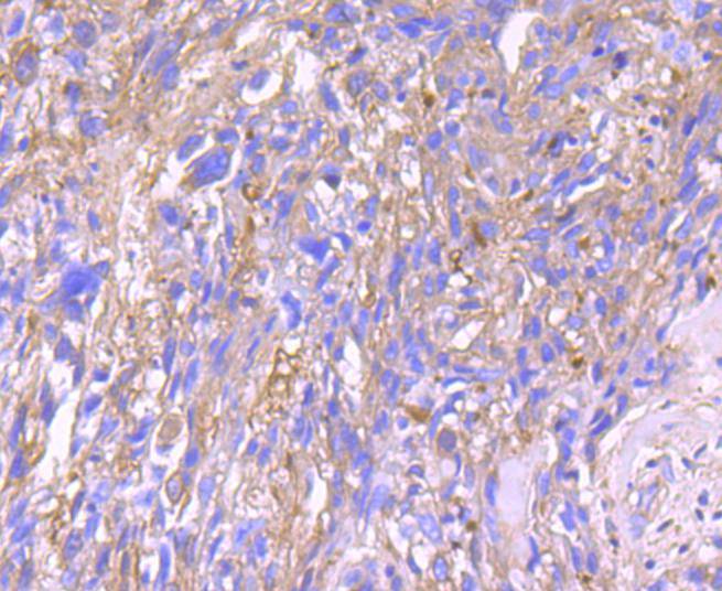
Immunohistochemical analysis of paraffin-embedded human breast carcinoma tissue using anti-alpha 1 Antitrypsin antibody. Counter stained with hematoxylin.
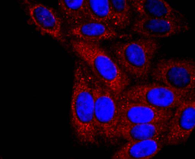
ICC staining alpha 1 Antitrypsin in Hela cells (red). The nuclear counter stain is DAPI (blue). Cells were fixed in paraformaldehyde, permeabilised with 0.25% Triton X100/PBS.
ICC staining alpha 1 Antitrypsin in MCF-7 cells (red). The nuclear counter stain is DAPI (blue). Cells were fixed in paraformaldehyde, permeabilised with 0.25% Triton X100/PBS.
ICC staining alpha 1 Antitrypsin in HepG2 cells (red). The nuclear counter stain is DAPI (blue). Cells were fixed in paraformaldehyde, permeabilised with 0.25% Triton X100/PBS.
Cumulative damage to lung tissue by Neutrophil Elastase is responsible for the development of pulmonary emphysema, an irreversible lung disease characterized by loss of lung elasticity. a 1-antitrypsin (AAT), a 394 amino acid hepatic acute phase protein, predominantly inhibits Neutrophil Elastase. AAT is highly expressed in liver and in cultured hepatoma cells and, to a lesser extent, in macrophages. AAT is a highly polymorphic glycosylated serum protein with characteristic isoelectric-focusing patterns for most variants. The gene encoding AAT maps to a region of human chromosome 14 that includes a related serine protease inhibitor (serpin) gene which encodes corticosteroid-binding globulin. Oxidation of the methionine 358 residue in the active center of AAT results in a dramatic decrease in inhibitory activity towards elastase. AAT also has a moderate affinity for plasmin and Thrombin. AAT deficiency is associated with a 20-30 fold increased risk of precocious pulmonary emphysema.
If you have published an article using product 49401, please notify us so that we can cite your literature.


 Yes
Yes


