Product Detail
Product NameTHY1 Rabbit mAb
Clone No.JF10-09
Host SpeciesRecombinant Rabbit
Clonality Monoclonal
PurificationProA affinity purified
ApplicationsWB, ICC/IF, IHC
Species ReactivityHu
Immunogen Descrecombinant protein
ConjugateUnconjugated
Other NamesCD7 antibody CD90 antibody CD90 antigen antibody CDw90 antibody FLJ33325 antibody MGC128895 antibody T25 antibody Theta antigen antibody Thy 1 antibody Thy 1 cell surface antigen antibody Thy 1 membrane glycoprotein antibody Thy 1 T cell antigen antibody Thy 1.2 antibody Thy-1 antigen antibody Thy-1 membrane glycoprotein antibody Thy1 antibody Thy1 antigen antibody Thy1 T cell antigen antibody Thy1.1 antibody Thy1.2 antibody THY1_HUMAN antibody Thymus cell antigen 1, theta antibody
Accession NoSwiss-Prot#:P04216
Uniprot
P04216
Gene ID
7070;
Calculated MW45 kDa
Formulation1*TBS (pH7.4), 1%BSA, 40%Glycerol. Preservative: 0.05% Sodium Azide.
StorageStore at -20˚C
Application Details
WB: 1:1,000-5,000
IHC: 1:50-1:200
ICC: 1:50-1:200
Western blot analysis of THY1 on different lysates using anti-THY1 antibody at 1/1,000 dilution. Positive control: Lane 1: Hela Lane 2: HepG2 Lane 3: HUVEC
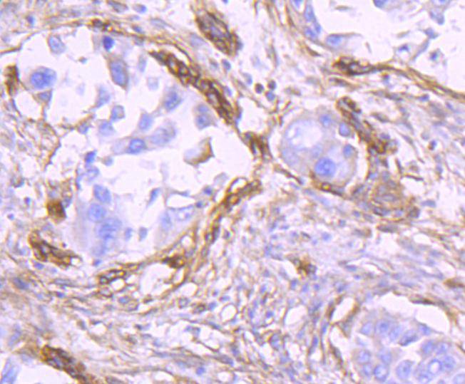
Immunohistochemical analysis of paraffin-embedded human lung cancer tissue using anti-THY1 antibody. Counter stained with hematoxylin.
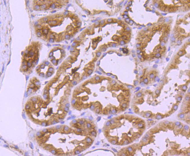
Immunohistochemical analysis of paraffin-embedded human kidney tissue using anti-THY1 antibody. Counter stained with hematoxylin.
Immunohistochemical analysis of paraffin-embedded human tonsil tissue using anti-THY1 antibody. Counter stained with hematoxylin.
Immunohistochemical analysis of paraffin-embedded human spleen tissue using anti-THY1 antibody. Counter stained with hematoxylin.
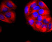
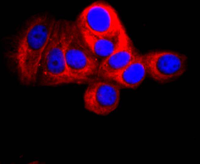
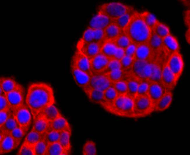
ICC staining THY1 in Hela cells (red). The nuclear counter stain is DAPI (blue). Cells were fixed in paraformaldehyde, permeabilised with 0.25% Triton X100/PBS.
ICC staining THY1 in MCF-7 cells (red). The nuclear counter stain is DAPI (blue). Cells were fixed in paraformaldehyde, permeabilised with 0.25% Triton X100/PBS.
ICC staining THY1 in PC-12 cells (red). The nuclear counter stain is DAPI (blue). Cells were fixed in paraformaldehyde, permeabilised with 0.25% Triton X100/PBS.
Over 100 cell surface markers have been identified through the use of monoclonal antibodies. Many of these markers have proven useful in identifying specific subpopulations of cells within mixed colonies. Accordingly, these molecules have been assigned a "cluster of differentiation" (CD) designation. One such marker, designated Thy-1 (also referred to as CDw90), is a phosphatidyl-anchored cell surface glycoprotein which, when coexpressed with CD34 on cells from normal human bone marrow, identifies a subpopulation that includes putative hematopoietic, pleuripotent stem cells. Thy-1+ cells from bone marrow have been implicated in syngeneic graft versus host disease and may serve to regulate autoreactivity after bone marrow transplant.
If you have published an article using product 49406, please notify us so that we can cite your literature.




 Yes
Yes


