Product Detail
Product NameVitamin D Binding protein Rabbit mAb
Clone No.JM10-36
Host SpeciesRecombinant Rabbit
Clonality Monoclonal
PurificationProA affinity purified
ApplicationsWB, ICC/IF, IHC, FC
Species ReactivityHu
Immunogen Descrecombinant protein
ConjugateUnconjugated
Other NamesDBP antibody
DBP/GC antibody
GC antibody
Gc globulin antibody
Gc-globulin antibody
GRD3 antibody
Group specific component antibody
Group specific component vitamin D binding protein antibody
Group-specific component antibody
hDBP antibody
VDB antibody
VDBG antibody
VDBP antibody
Vitamin D binding alpha globulin antibody
Vitamin D-binding protein antibody
VTDB_HUMAN antibody
Accession NoSwiss-Prot#:P02774
Uniprot
P02774
Gene ID
2638;
Calculated MW53 kDa
Formulation1*TBS (pH7.4), 1%BSA, 40%Glycerol. Preservative: 0.05% Sodium Azide.
StorageStore at -20˚C
Application Details
WB: 1:1,000-1:2,000
IHC: 1:50-1:200
ICC: 1:50-1:200
FC: 1:50-1:100
Western blot analysis of DBP on human lung lysates using anti- DBP antibody at 1/1,000 dilution.
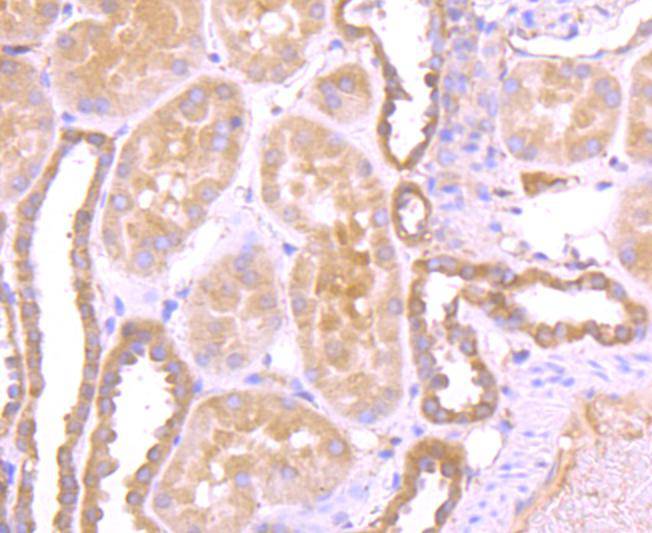
Immunohistochemical analysis of paraffin-embedded human kidney tissue using anti- DBP antibody. Counter stained with hematoxylin.
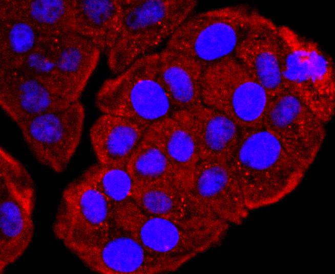
ICC staining DBP in Hela cells (red). The nuclear counter stain is DAPI (blue). Cells were fixed in paraformaldehyde, permeabilised with 0.25% Triton X100/PBS.
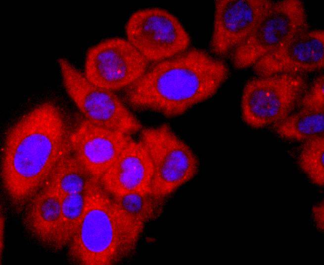
ICC staining DBP in HepG2 cells (red). The nuclear counter stain is DAPI (blue). Cells were fixed in paraformaldehyde, permeabilised with 0.25% Triton X100/PBS.
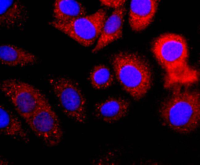
ICC staining DBP in SKOV-3 cells (red). The nuclear counter stain is DAPI (blue). Cells were fixed in paraformaldehyde, permeabilised with 0.25% Triton X100/PBS.
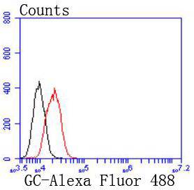
Flow cytometric analysis of HepG2 cells with DBP antibody at 1/50 dilution (red) compared with an unlabelled control (cells without incubation with primary antibody; black). Alexa Fluor 488-conjugated goat anti rabbit IgG was used as the secondary antibody.
Vitamin D-binding protein (DBP) is a multi-functional serum protein that binds to the plasma membranes of numerous cell types and mediates a variety of cellular functions. The locus of the DBP protein (also known as group-specific component protein or GC) is located at human chromosome 4q13.3. DBP functions in organ-specific transportation of vitamin D and its metabolites to the various target organs of the vitamin D endocrine system. In addition, DBP has immunomodulatory properties and is able to bind to the surface of leukocytes. DBP binds to the plasma membrane through a chondroitin sulfate proteoglycan. DBP serves as a co-chemotactic factor for C5a to enhance the chemotactic activity of C5a. DBP can also bind to globular Actin with high affinity and is involved in the clearance of Actin from the blood. DBP plays an important role in osteoclast differentiation. The diverse cellular functions of DBP require its cell surface binding ability to mediate different biological processes.
If you have published an article using product 49422, please notify us so that we can cite your literature.


 Yes
Yes


