Welcome to Sabbiotech!     
Email:info@signalwayantibody.com
Location: Home > Recombinant Rabbit Monoclonal Antibodies > CD40L Rabbit mAb

CD40L Rabbit mAb#49477

Availability: Yes
Total Amount:$
Product Detail

Product NameCD40L Rabbit mAb

Clone No.SR4543

Host SpeciesRecombinant Rabbit

Clonality Monoclonal

PurificationProA affinity purified

ApplicationsWB, ICC/IF,IHC, FC

Species ReactivityHu, Ms,Rt

Immunogen Descrecombinant protein

ConjugateUnconjugated

Other NamesCD 40L antibody
CD154 antibody
CD40 antigen ligand antibody
CD40 ligand antibody
CD40 ligand, soluble form antibody
CD40-L antibody
CD40L antibody
CD40L_HUMAN antibody
CD40LG antibody
gp39 antibody
hCD40L antibody
HIGM1 antibody
IGM antibody
IMD3 antibody
T B cell activating molecule antibody
T BAM antibody
T-cell antigen Gp39 antibody
TNF-related activation protein antibody
TNFSF5 antibody
TrAP antibody
Tumor necrosis factor (ligand) superfamily member 5 antibody
Tumor necrosis factor ligand superfamily member 5 antibody

Accession NoSwiss-Prot#:P29965

Uniprot P29965

Gene ID 959;

Calculated MW36 kDa

Formulation1*TBS (pH7.4), 1%BSA, 40%Glycerol. Preservative: 0.05% Sodium Azide.

StorageStore at -20˚C

Application Details

WB: 1:500-1:1,000
IHC: 1:50-1:200
ICC: 1:50-1:200

FC: 1:50-1:100

Western blot analysis of CD40L on different cells lysates using anti-CD40L antibody at 1/500 dilution. Positive control: Lane 1: Hela Lane 2: Mouse liver
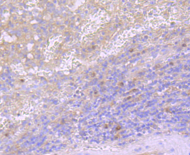
Immunohistochemical analysis of paraffin-embedded human spleen tissue using anti- CD40L antibody. Counter stained with hematoxylin.
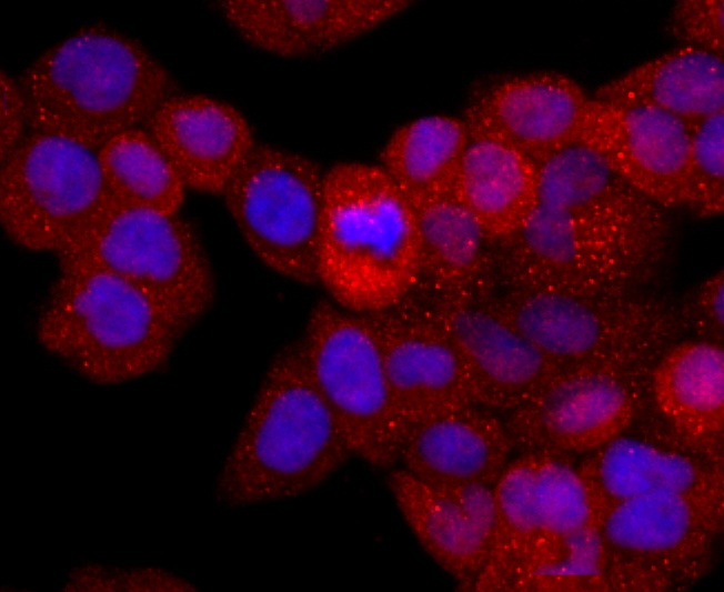
ICC staining CD40L in Hela cells (red). The nuclear counter stain is DAPI (blue). Cells were fixed in paraformaldehyde, permeabilised with 0.25% Triton X100/PBS.
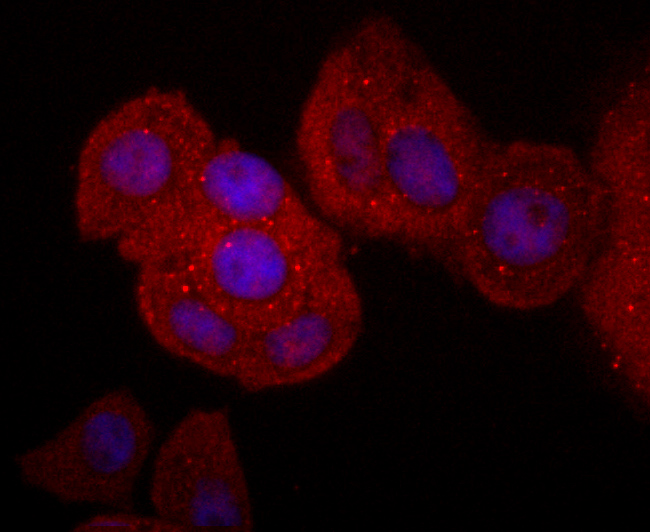
ICC staining CD40L in MCF-7 cells (red). The nuclear counter stain is DAPI (blue). Cells were fixed in paraformaldehyde, permeabilised with 0.25% Triton X100/PBS.
ICC staining CD40L in NIH-3T3 cells (green). The nuclear counter stain is DAPI (blue). Cells were fixed in paraformaldehyde, permeabilised with 0.25% Triton X100/PBS.
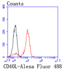
Flow cytometric analysis of THP-1 cells with CD40L antibody at 1/50 dilution (red) compared with an unlabelled control (cells without incubation with primary antibody; black). Alexa Fluor 488-conjugated goat anti rabbit IgG was used as the secondary antibody.

Resting B cells can be activated and clonally expanded into antibody-producing cells in response to a combination of cell contact and soluble signals provided by primed helper T (Th) cells. While cytokines IL-4 and IL-13 alone are inadequate for B cell activation, contact with Th cells seems to be sufficient for delivery of proliferative signals. CD40 and CD154 (also designated CD40L or TRAP) comprise a receptor ligand pair central to the transmission of this signal. CD40 is expressed on the surface of B cells and CD154 is expressed on activated T cells. In the presence of such stimulus, IL-4 and IL-13 are capable of triggering immunoglobulin class switching and secretion of IgE. CD154 is a 261 amino acid protein that is is expressed as a soluble cytokine as well as a homotrimeric type II transmembrane protein. Its expression is tightly regulated, and abnormal levels of CD154 are associated with the pathogenesis of atheromatous plaque destabilization and thrombotic events. Mutations in the gene encoding for CD154 are implicated in hyper-IgM immunodeficiency syndrome type 1.

If you have published an article using product 49477, please notify us so that we can cite your literature.

NOTE

Application

  • WBWestern Blotting
  • IHCImmunohistochemistry
  • IFImmunofluorescence
  • ICCImmunocytochemistry
  • FCFlow Cytometry
  • IPImmunoprecipitation
  • EELISA
  • DBDot Blotting
  • ChIPChromatin Immunoprecipitation
  • GICAGold Immunochromatography Assay
  • NCNegative Control

Species Reactivity

  • HuHuman
  • MsMouse
  • RtRat
  • DmDrosophila melanogaster
  • CCaenorhabditis elegans
  • MkMonkey
  • RbRabbit
  • BBovine
  • DDog
  • PPig
  • HmHamster
  • ChHmChinese Hamster
  • ChkChicken
  • ShpSheep
;