Welcome to Sabbiotech!     
Email:info@signalwayantibody.com
Location: Home > Recombinant Rabbit Monoclonal Antibodies > TDP43 Rabbit mAb

TDP43 Rabbit mAb#49487

Availability: Yes
Total Amount:$
Product Detail

Product NameTDP43 Rabbit mAb

Clone No.JM51-10

Host SpeciesRecombinant Rabbit

Clonality Monoclonal

PurificationProA affinity purified

ApplicationsWB, IP, ICC/IF, IHC, FC

Species ReactivityHu, Ms, Rt, Zebrafish

Immunogen Descrecombinant protein

ConjugateUnconjugated

Other NamesALS10 antibody
OTTHUMP00000002171 antibody
OTTHUMP00000002172 antibody
OTTHUMP00000002173 antibody
TADBP_HUMAN antibody
TAR DNA binding protein 43 antibody
TAR DNA binding protein antibody
TAR DNA-binding protein 43 antibody
TARDBP antibody
TDP 43 antibody
TDP-43 antibody
TDP43 antibody

Accession NoSwiss-Prot#:Q13148

Uniprot Q13148

Gene ID 23435;

Calculated MW45 kDa

Formulation1*TBS (pH7.4), 1%BSA, 40%Glycerol. Preservative: 0.05% Sodium Azide.

StorageStore at -20˚C

Application Details
WB: 1:1,000-5,000
IHC: 1:50-1:200
ICC: 1:100-1:500
IP: 1:10-1:50
FC: 1:50-1:100
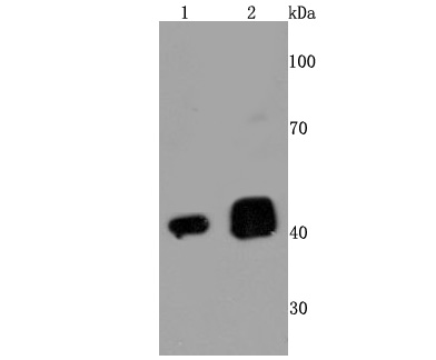
Western blot analysis of TDP43 on different cells lysates using anti-TDP43 antibody at 1/500 dilution. Positive control��
Line 1: Hela
Line 2: K563
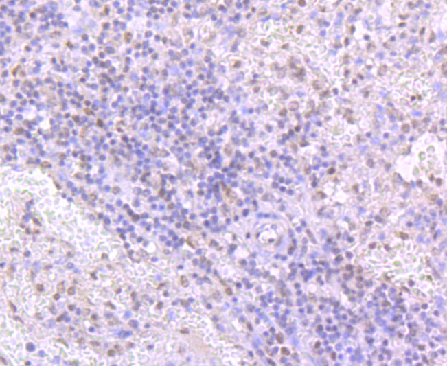
Immunohistochemical analysis of paraffin-embedded human spleen tissue using anti-TDP43 antibody. Counter stained with hematoxylin.
Immunohistochemical analysis of paraffin-embedded human pancreas tissue using anti-TDP43 antibody. Counter stained with hematoxylin.
Immunohistochemical analysis of paraffin-embedded mouse placenta tissue using anti-TDP43 antibody. Counter stained with hematoxylin.
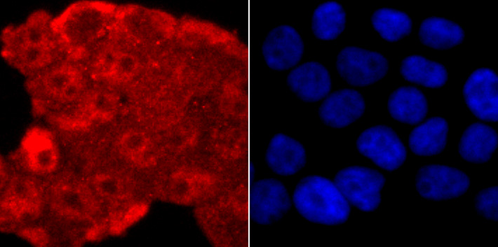
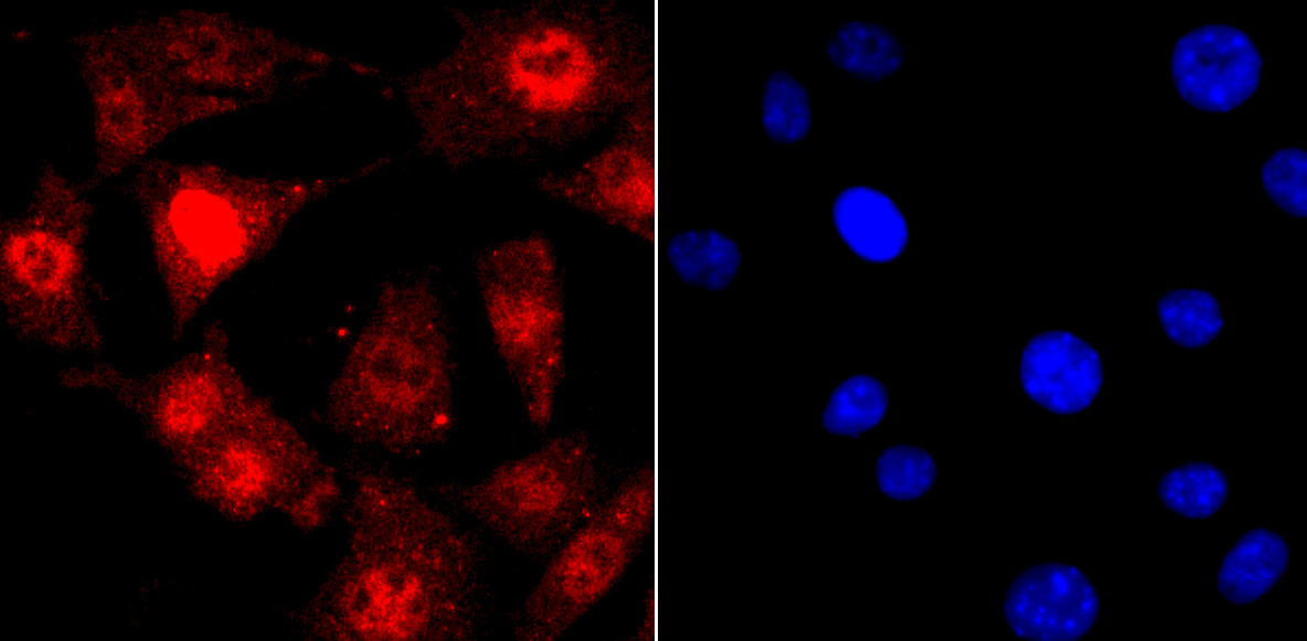
ICC staining TDP43 in 293T cells (red). The nuclear counter stain is DAPI (blue). Cells were fixed in paraformaldehyde, permeabilised with 0.25% Triton X100/PBS.
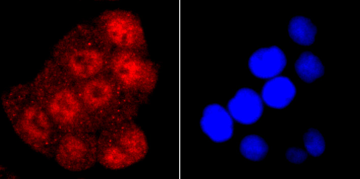
ICC staining TDP43 in Hela cells (red). The nuclear counter stain is DAPI (blue). Cells were fixed in paraformaldehyde, permeabilised with 0.25% Triton X100/PBS.
ICC staining TDP43 in SH-SY5Y cells (red). The nuclear counter stain is DAPI (blue). Cells were fixed in paraformaldehyde, permeabilised with 0.25% Triton X100/PBS.
Flow cytometric analysis of Hela cells with TDP43 antibody at 1/50 dilution (red) compared with an unlabelled control (cells without incubation with primary antibody; black). Alexa Fluor 488-conjugated goat anti rabbit IgG was used as the secondary antibody.
DNA and RNA-binding protein which regulates transcription and splicing. Involved in the regulation of CFTR splicing. It promotes CFTR exon 9 skipping by binding to the UG repeated motifs in the polymorphic region near the 3'-splice site of this exon. The resulting aberrant splicing is associated with pathological features typical of cystic fibrosis. May also be involved in microRNA biogenesis, apoptosis and cell division. Can repress HIV-1 transcription by binding to the HIV-1 long terminal repeat. Stabilizes the low molecular weight neurofilament (NFL) mRNA through a direct interaction with the 3' UTR.

If you have published an article using product 49487, please notify us so that we can cite your literature.

NOTE

Application

  • WBWestern Blotting
  • IHCImmunohistochemistry
  • IFImmunofluorescence
  • ICCImmunocytochemistry
  • FCFlow Cytometry
  • IPImmunoprecipitation
  • EELISA
  • DBDot Blotting
  • ChIPChromatin Immunoprecipitation
  • GICAGold Immunochromatography Assay
  • NCNegative Control

Species Reactivity

  • HuHuman
  • MsMouse
  • RtRat
  • DmDrosophila melanogaster
  • CCaenorhabditis elegans
  • MkMonkey
  • RbRabbit
  • BBovine
  • DDog
  • PPig
  • HmHamster
  • ChHmChinese Hamster
  • ChkChicken
  • ShpSheep
;