Welcome to Sabbiotech!     
Email:info@signalwayantibody.com
Location: Home > Recombinant Rabbit Monoclonal Antibodies > Annexin A2 Rabbit mAb

Annexin A2 Rabbit mAb#49559

Availability: Yes
Total Amount:$
Product Detail

Product NameAnnexin A2 Rabbit mAb

Clone No.JA42-30

Host SpeciesRecombinant Rabbit

Clonality Monoclonal

PurificationProA affinity purified

ApplicationsWB, IHC, ICC/IF, FC

Species ReactivityHu, Ms, Rt

Immunogen Descrecombinant protein

ConjugateUnconjugated

Other NamesAnnexin A2 antibody
Annexin II antibody
Annexin II, heavy chain antibody
Annexin-2 antibody
ANX 2 antibody
ANX2 antibody
ANX2L4 antibody
ANXA2 antibody
ANXA2_HUMAN antibody
arylsulfatase B antibody
CAL1H antibody
Calpactin I heavy chain antibody
calpactin I heavy polypeptide (p36) antibody
Calpactin I heavy polypeptide antibody
Calpactin-1 heavy chain antibody
chromobindin 8 antibody
Chromobindin-8 antibody
Epididymis secretory protein Li 270 antibody
HEL S 270 antibody
LIP2 antibody
Lipocortin II antibody
LPC2 antibody
LPC2D antibody
p36 antibody
P36 protein antibody
PAP-IV antibody
Placental anticoagulant protein IV antibody
Protein I antibody

Accession NoSwiss-Prot#:P07355

Uniprot P07355

Gene ID 302;

Calculated MW38 kDa

Formulation1*TBS (pH7.4), 1%BSA, 40%Glycerol. Preservative: 0.05% Sodium Azide.

StorageStore at -20˚C

Application Details
WB: 1:500-1:2,000
IHC: 1:50-1:200
ICC:1:100-1:200

FC:1:50-1:200
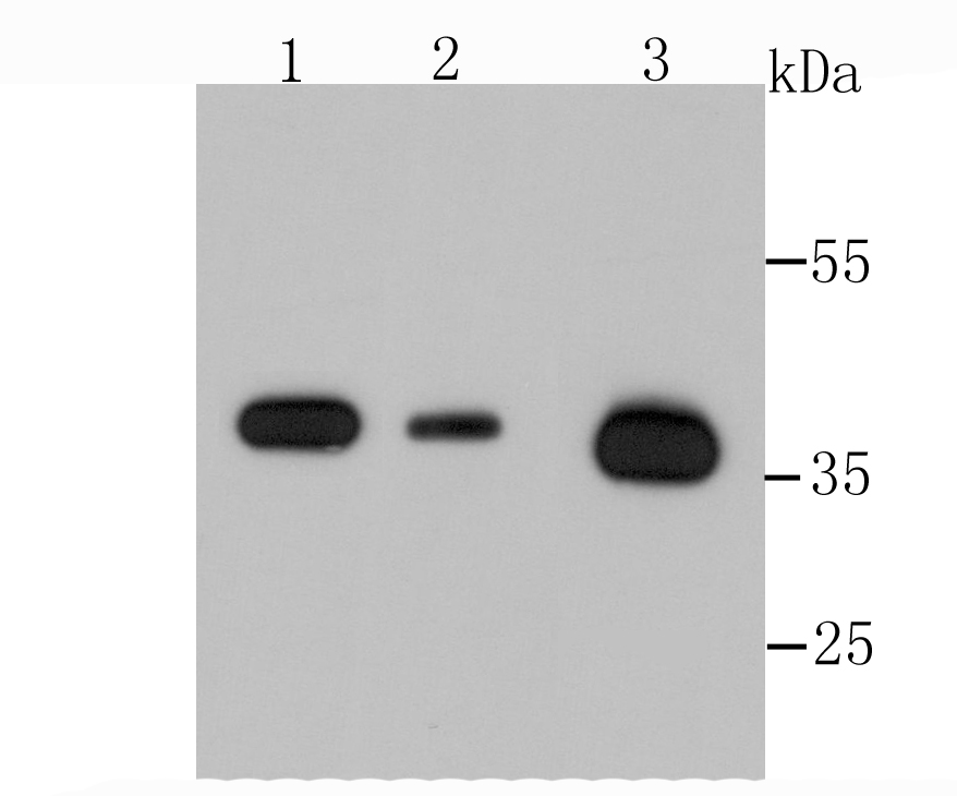
Western blot analysis of Annexin A2 on different cells lysates using anti-Annexin A2 antibody at 1/500 dilution. Positive control��
Lane 1: Hela
Lane 2: Human kidney
Lane 3: Murine testis
Immunohistochemical analysis of paraffin-embedded human liver tissue using anti-Annexin A2 antibody. Counter stained with hematoxylin.
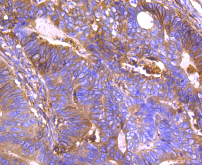
Immunohistochemical analysis of paraffin-embedded human colon cancer tissue using anti-Annexin A2 antibody. Counter stained with hematoxylin.
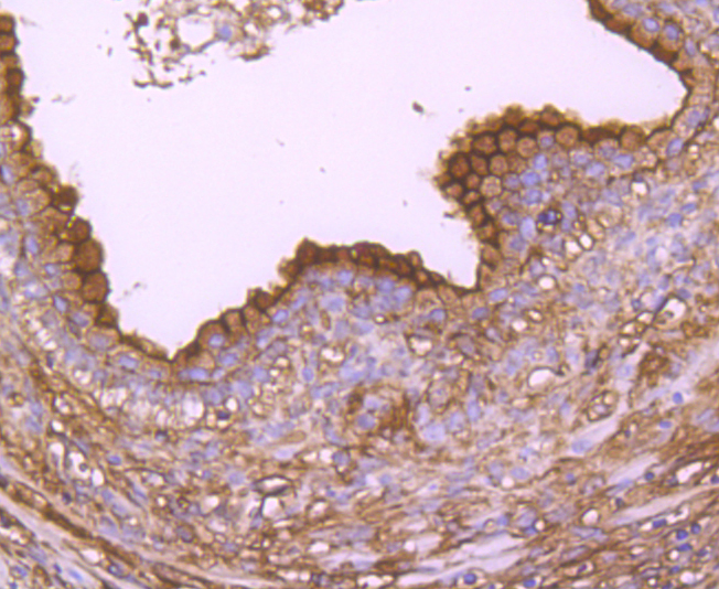
Immunohistochemical analysis of paraffin-embedded human prostate tissue using anti-Annexin A2 antibody. Counter stained with hematoxylin.
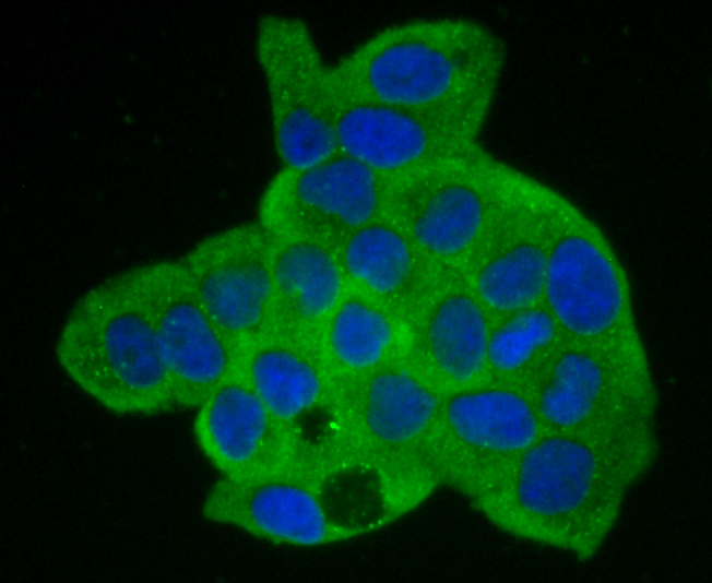
ICC staining Annexin A2 in Hela cells (green). The nuclear counter stain is DAPI (blue). Cells were fixed in paraformaldehyde, permeabilised with 0.25% Triton X100/PBS.
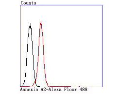
Flow cytometric analysis of K562 cells with Annexin A2 antibody at 1/100 dilution (red) compared with an unlabelled control (cells without incubation with primary antibody; black).
The annexin family of calcium-binding proteins is composed of at least ten mammalian genes and is characterized by a conserved core domain which binds phospholipids in a Ca2+-dependent manner and a unique amino-terminal region which may confer binding specificity. The interaction between these proteins and biological membranes has led to the hypothesis that they are involved in cellular trafficking processes such as endocytosis, exocytosis and cellular adhesion. Annexin I, alternatively referred to as lipocortin, has been implicated as a mediator of the anti-inflammatory response produced by glucocorticoids and as an inhibitor of cPLA2, a potent mediator of inflammation. Annexin II, also called p36, exists as a monomer or as a heterotetramer, complexed with the S-100-related protein p11. This complex is termed calpactin I. In the tetrameric form, Annexin II is an efficient substrate of PKC family and Src pp60.

If you have published an article using product 49559, please notify us so that we can cite your literature.

NOTE

Application

  • WBWestern Blotting
  • IHCImmunohistochemistry
  • IFImmunofluorescence
  • ICCImmunocytochemistry
  • FCFlow Cytometry
  • IPImmunoprecipitation
  • EELISA
  • DBDot Blotting
  • ChIPChromatin Immunoprecipitation
  • GICAGold Immunochromatography Assay
  • NCNegative Control

Species Reactivity

  • HuHuman
  • MsMouse
  • RtRat
  • DmDrosophila melanogaster
  • CCaenorhabditis elegans
  • MkMonkey
  • RbRabbit
  • BBovine
  • DDog
  • PPig
  • HmHamster
  • ChHmChinese Hamster
  • ChkChicken
  • ShpSheep
;