Welcome to Sabbiotech!     
Email:info@signalwayantibody.com
Location: Home > Recombinant Rabbit Monoclonal Antibodies > APE1 Rabbit mAb

APE1 Rabbit mAb#59488

Availability: Yes
Total Amount:$
Product Detail

Product NameAPE1 Rabbit mAb

Host SpeciesRabbit

ClonalityMonoclonal

IsotypeRabbit IgG

PurificationAffinity-chromatography

ApplicationsWB IHC ICC/IF

Species ReactivityHuman Mouse Rat

SpecificityAPE1 Antibody detects endogenous levels of total APE1

Immunogen DescA synthesized peptide derived from human APE1

Other NamesDNA-(apurinic or apyrimidinic site) lyase; APEX nuclease; APEN; AP endonuclease 1; APE-1; APEX1; APE; APE1; APEX; APX; HAP1; REF1;

Accession NoUniprot:P27695

Uniprot P27695

Calculated MWPredicted band size: 36 kDa

Sdspage MWObserved band size: 36 kDa

FormulationRabbit IgG in phosphate buffered saline , pH 7.4, 150mM NaCl, 0.02% sodium azide and 50% glycerol.

StorageStore at +4˚C short term. Store at -20˚C long term. Avoid freeze / thaw cycle.

Application Details
WB: 1:500-1:2000
IHC: 1:50-1:200
ICC/IF: 1:50-1:200
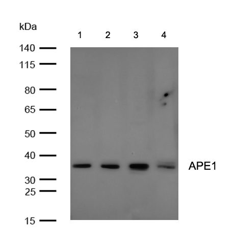
All lanes: APE1 Rabbit mAb at 1/1k dilution

Lane 1 : K562 whole cell lysates
Lane 2 : Hela whole cell lysates
Lane 3 : 3T3 whole cell lysates
Lane 4 : C6 whole cell lysates
Lysates/proteins at 20 µg per lane.

Secondary
All lanes : Goat Anti-Rabbit IgG H&L (HRP) at 1/20000 dilution

Predicted band size: 36 kDa
Observed band size: 36 kDa

Exposure time: 7 seconds
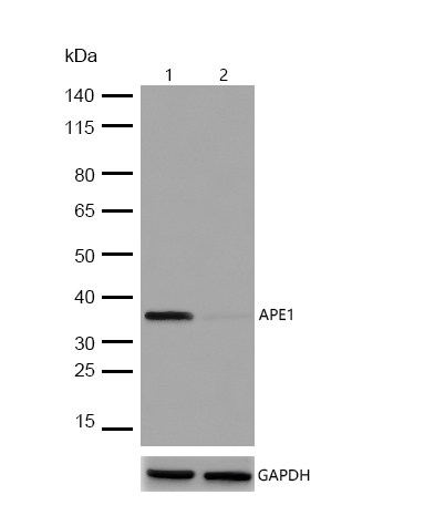
All lanes:APE1 Rabbit mAb at 1/1k dilution

Lane 1 : Wild-type A549 cell lysate
Lane 2 :APE1 Rabbit mAb knockdown A549 cell lysate

Lysates/proteins at 20 µg per lane.
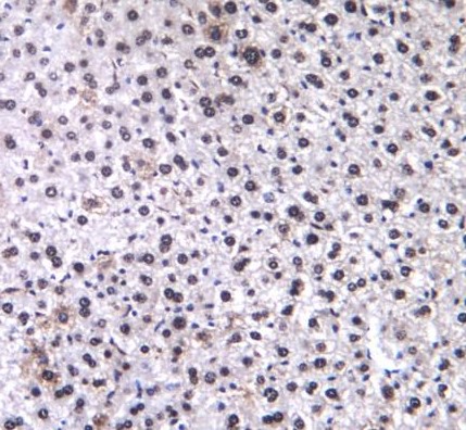
Formalin-fixed, paraffin-embedded mouse liver tissue stained for APE1 using 59488 at 1/100 dilution in immunohistochemical analysis.
Immunocytochemistry/ Immunofluorescence APE1 antibody (59488)

ICC/IF staining of APE1 in Hela cells. Cells were fixed with 4% Paraformaldehyde permeabilized with 0.1% Triton X-100.
Samples were incubated with 59488 at a working dilution of 1/100. The secondary antibody was Alexa Fluor® 488 goat anti rabbit, used at a dilution of 1/500.
Nuclei were counterstained with DAPI.
Ape1 initiates the repair of abasic sites and is essential for the base excision repair (BER) pathway. Repair activities of Ape1 are stimulated by interaction with XRCC1, another essential protein in BER. Ape1 functions as a redox factor that maintains transcription factors in an active, reduced state but can also function in a redox-independent manner as a transcriptional cofactor to control different cellular fates such as apoptosis, proliferation and differentiation.

If you have published an article using product 59488, please notify us so that we can cite your literature.

NOTE

Application

  • WBWestern Blotting
  • IHCImmunohistochemistry
  • IFImmunofluorescence
  • ICCImmunocytochemistry
  • FCFlow Cytometry
  • IPImmunoprecipitation
  • EELISA
  • DBDot Blotting
  • ChIPChromatin Immunoprecipitation
  • GICAGold Immunochromatography Assay
  • NCNegative Control

Species Reactivity

  • HuHuman
  • MsMouse
  • RtRat
  • DmDrosophila melanogaster
  • CCaenorhabditis elegans
  • MkMonkey
  • RbRabbit
  • BBovine
  • DDog
  • PPig
  • HmHamster
  • ChHmChinese Hamster
  • ChkChicken
  • ShpSheep
;