Product Detail
Product NameMyD88 Rabbit mAb
Clone No.SC65-04
Host SpeciesRecombinant Rabbit
Clonality Monoclonal
PurificationProA affinity purified
ApplicationsWB, ICC/IF, IHC, FC
Species ReactivityHu
Immunogen Descrecombinant protein
ConjugateUnconjugated
Other NamesMutant myeloid differentiation primary response 88 antibody MYD 88 antibody Myd88 antibody MYD88_HUMAN antibody MYD88D antibody Myeloid differentiation marker 88 antibody Myeloid differentiation primary response 88 antibody Myeloid differentiation primary response gene (88) antibody Myeloid differentiation primary response gene 88 antibody Myeloid differentiation primary response gene antibody Myeloid differentiation primary response protein MyD88 antibody OTTHUMP00000161718 antibody OTTHUMP00000208595 antibody OTTHUMP00000209058 antibody OTTHUMP00000209059 antibody OTTHUMP00000209060 antibody
Accession NoSwiss-Prot#:Q99836
Uniprot
Q99836
Gene ID
4615;
Calculated MW33 kDa
Formulation1*TBS (pH7.4), 1%BSA, 40%Glycerol. Preservative: 0.05% Sodium Azide.
StorageStore at -20˚C
Application Details
WB: 1:1,000
IHC: 1:50-1:200
ICC: 1:100-1:500
FC: 1:50-1:100
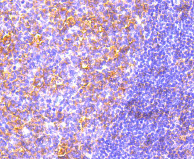
Immunohistochemical analysis of paraffin-embedded human tonsil tissue using anti-MyD88 antibody. Counter stained with hematoxylin.
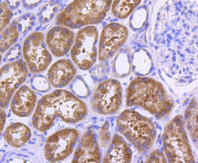
Immunohistochemical analysis of paraffin-embedded human kidney tissue using anti-MyD88 antibody. Counter stained with hematoxylin.
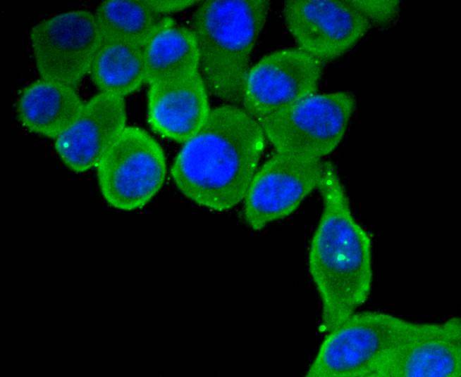
ICC staining MyD88 in HepG2 cells (green). The nuclear counter stain is DAPI (blue). Cells were fixed in paraformaldehyde, permeabilised with 0.25% Triton X100/PBS.
ICC staining MyD88 in A549 cells (green). The nuclear counter stain is DAPI (blue). Cells were fixed in paraformaldehyde, permeabilised with 0.25% Triton X100/PBS.
ICC staining MyD88 in MCF-7 cells (green). The nuclear counter stain is DAPI (blue). Cells were fixed in paraformaldehyde, permeabilised with 0.25% Triton X100/PBS.
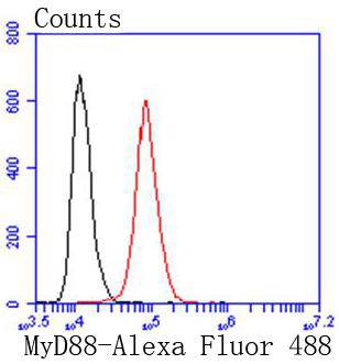
Flow cytometric analysis of Hela cells with MyD88 antibody at 1/50 dilution (red) compared with an unlabelled control (cells without incubation with primary antibody; black). Alexa Fluor 488-conjugated goat anti rabbit IgG was used as the secondary antibody
Interleukin-1 (IL-1)-induced activation of the NFκB pathway is mediated through the IL-1 receptor and the subsequent phosphorylation of IL-1 receptor-associated kinase (IRAK). The myeloid differentiation protein MyD88 was originally characterized as a protein upregulated in myeloleukemic cells following IL-6-induced growth arrest and terminal differentiation. MyD88 is now known to function as an adaptor protein for the association of IRAK with the IL-1 receptor. MyD88 is functionally homologous to the adaptor protein tube in the Toll signaling pathway of Drosophilia, and both proteins are members of the Toll/IL-1R superfamily. MyD88 contains a characteristic N-terminal death domain that is essential for NFκB activation and an adjacent Toll/IL-1R homology domain (TIR domain). Collectively, these domains enable the protein-protein interactions of MyD88 with IRAK and the IL-1 receptor complex.
If you have published an article using product 48999, please notify us so that we can cite your literature.


 Yes
Yes


